Copyright
©The Author(s) 2021.
World J Clin Cases. Mar 16, 2021; 9(8): 1893-1900
Published online Mar 16, 2021. doi: 10.12998/wjcc.v9.i8.1893
Published online Mar 16, 2021. doi: 10.12998/wjcc.v9.i8.1893
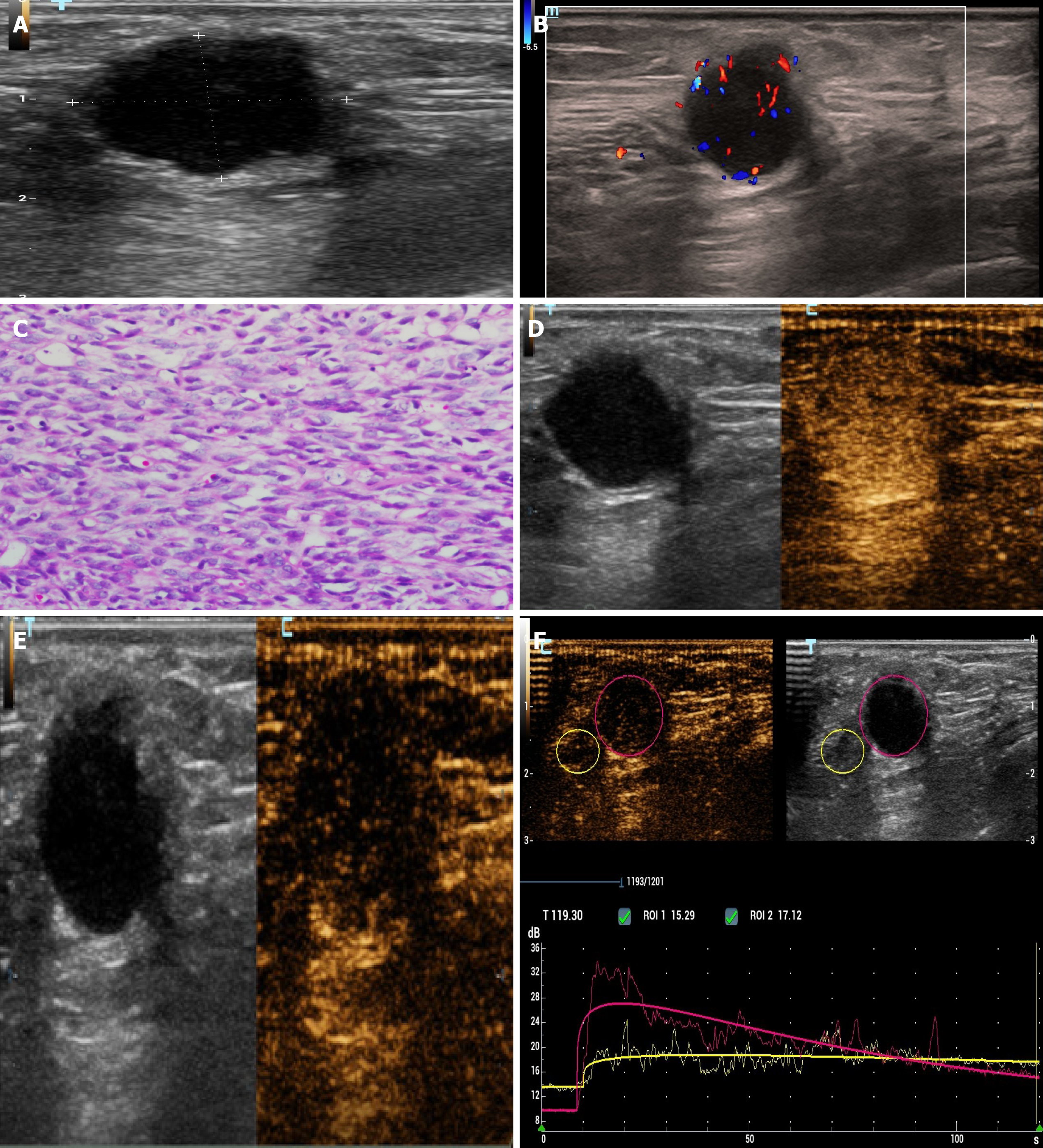
Figure 3 Chest wall metastasis.
A and B: The chest wall mass showed a hypoechoic mass in the superficial fascia layer on two-dimensional ultrasonography. Color Doppler flow imaging: Dot strip blood flow signal was seen in the mass; C: Pathological image of chest wall metastatic synovial sarcoma (hematoxylin and eosin staining, × 400); D-F: The ultrasound contrast agent quickly withdrew after rapidly high enhancement, showing the “fast forward and fast retreat” enhancement mode.
- Citation: Li R, Teng X, Han WH, Li Y, Liu QW. Imaging findings of primary pulmonary synovial sarcoma with secondary distant metastases: A case report. World J Clin Cases 2021; 9(8): 1893-1900
- URL: https://www.wjgnet.com/2307-8960/full/v9/i8/1893.htm
- DOI: https://dx.doi.org/10.12998/wjcc.v9.i8.1893


