Copyright
©The Author(s) 2021.
World J Clin Cases. Mar 6, 2021; 9(7): 1755-1760
Published online Mar 6, 2021. doi: 10.12998/wjcc.v9.i7.1755
Published online Mar 6, 2021. doi: 10.12998/wjcc.v9.i7.1755
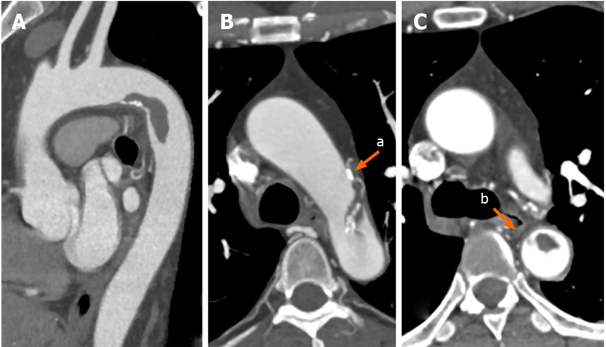
Figure 2 Preoperative computed tomography angiography.
A: The thrombus originated from the lesser curve of the aortic arch, and its distal part was floating; B: Calcified plaques and hematoma (arrow a) located at the aortic wall, and some calcified plaques were within the floating thrombus; and C: A pedicle (arrow b) connected the floating thrombus to the adjacent aortic wall.
- Citation: Wang TH, Zhao JC, Xiong F, Yang Y. Use of three dimensional-printing in the management of floating aortic thrombus due to occult aortic dissection: A case report. World J Clin Cases 2021; 9(7): 1755-1760
- URL: https://www.wjgnet.com/2307-8960/full/v9/i7/1755.htm
- DOI: https://dx.doi.org/10.12998/wjcc.v9.i7.1755


