Copyright
©The Author(s) 2021.
World J Clin Cases. Mar 6, 2021; 9(7): 1682-1695
Published online Mar 6, 2021. doi: 10.12998/wjcc.v9.i7.1682
Published online Mar 6, 2021. doi: 10.12998/wjcc.v9.i7.1682
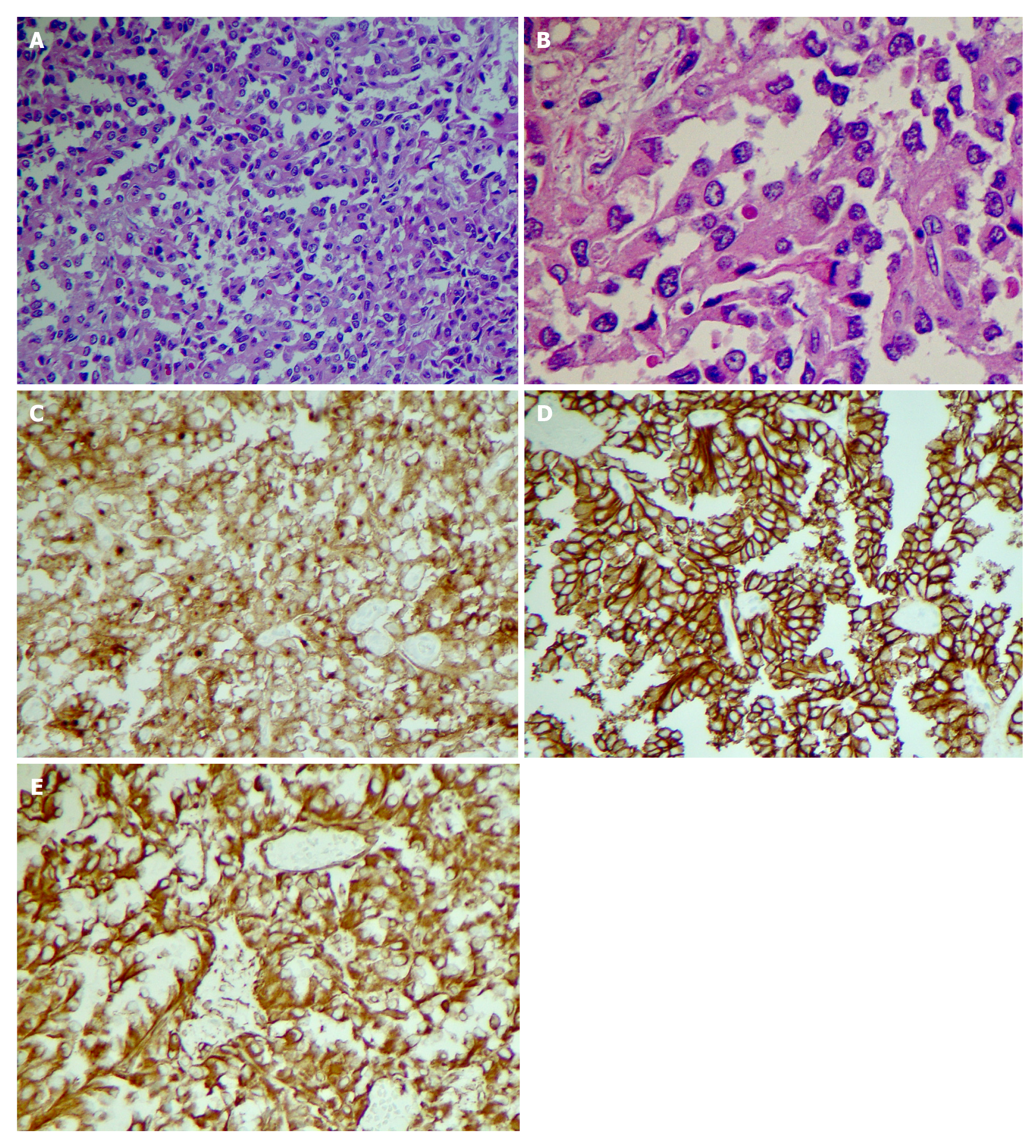
Figure 2 Pathological phenomena.
Histology shows solid areas with poorly cohesive cells forming cuff around blood vessels resulting in a pseudopapillary architecture. Tumor cells show uniform nuclei with finely textured chromatin, inconspicuous nucleoli and occasional longitudinal grooves. A: × 20 magnification; B: × 40 magnification; C: CD 10 positive; D: CD 56 positive; E: Vimentin positive.
- Citation: Abudalou M, Vega EA, Dhingra R, Holzwanger E, Krishnan S, Kondratiev S, Niakosari A, Conrad C, Stallwood CG. Solid pseudopapillary neoplasm-diagnostic approach and post-surgical follow up: Three case reports and review of literature. World J Clin Cases 2021; 9(7): 1682-1695
- URL: https://www.wjgnet.com/2307-8960/full/v9/i7/1682.htm
- DOI: https://dx.doi.org/10.12998/wjcc.v9.i7.1682


