Copyright
©The Author(s) 2021.
World J Clin Cases. Feb 26, 2021; 9(6): 1343-1352
Published online Feb 26, 2021. doi: 10.12998/wjcc.v9.i6.1343
Published online Feb 26, 2021. doi: 10.12998/wjcc.v9.i6.1343
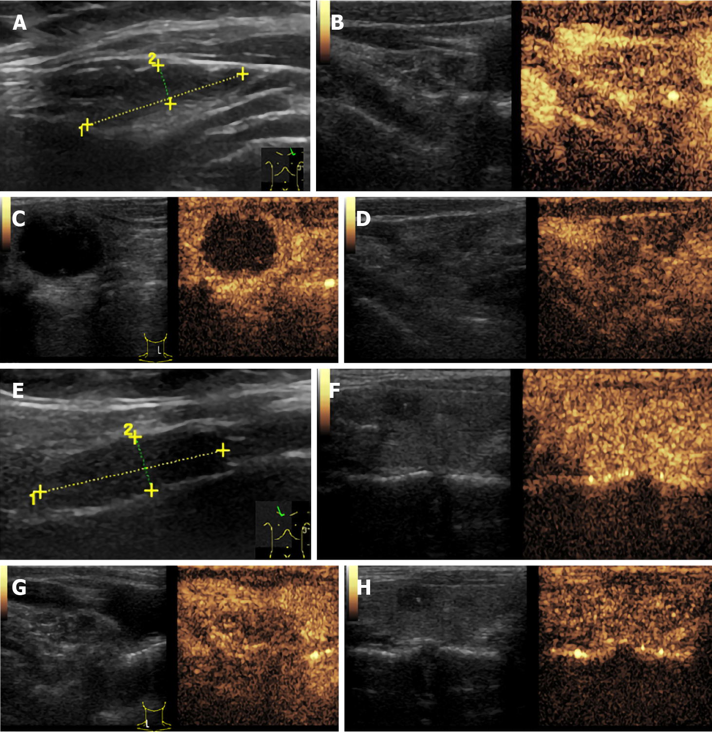
Figure 2 Ultrasound images.
A/E: Ultrasound suggested enlargement of cervical lymph nodes; B-D/F-H: Further contrast-enhanced ultrasound suggested uneven enhancement of enlarged lymph nodes from the medulla to cortex, with slightly lower medulla enhancement, suggesting that enlarged lymph nodes were mostly reactive hyperplasia.
- Citation: Gan FJ, Zhou T, Wu S, Xu MX, Sun SH. Do medullary thyroid carcinoma patients with high calcitonin require bilateral neck lymph node clearance? A case report. World J Clin Cases 2021; 9(6): 1343-1352
- URL: https://www.wjgnet.com/2307-8960/full/v9/i6/1343.htm
- DOI: https://dx.doi.org/10.12998/wjcc.v9.i6.1343


