Copyright
©The Author(s) 2021.
World J Clin Cases. Feb 26, 2021; 9(6): 1343-1352
Published online Feb 26, 2021. doi: 10.12998/wjcc.v9.i6.1343
Published online Feb 26, 2021. doi: 10.12998/wjcc.v9.i6.1343
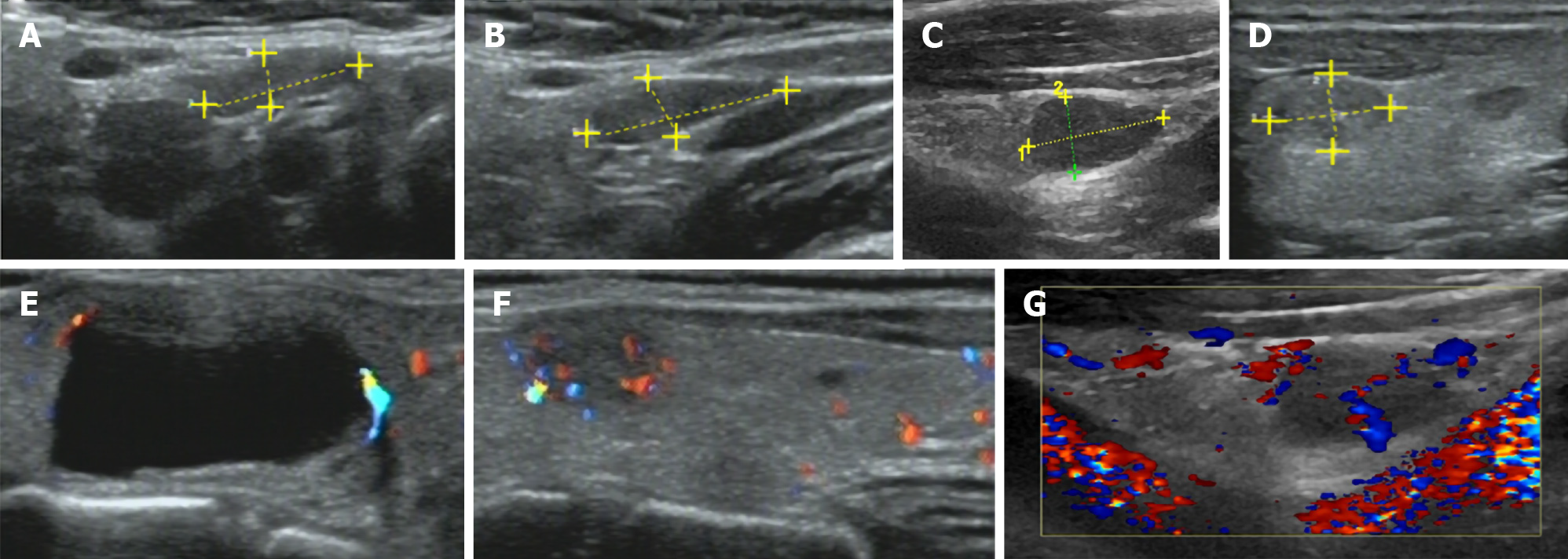
Figure 1 Ultrasound images.
A-D: Ultrasound (US) suggested multiple hypoechoic nodules in bilateral lobes of the thyroid, with clear boundaries; E-G: US indicated the dotted blood flow signal in nodules.
- Citation: Gan FJ, Zhou T, Wu S, Xu MX, Sun SH. Do medullary thyroid carcinoma patients with high calcitonin require bilateral neck lymph node clearance? A case report. World J Clin Cases 2021; 9(6): 1343-1352
- URL: https://www.wjgnet.com/2307-8960/full/v9/i6/1343.htm
- DOI: https://dx.doi.org/10.12998/wjcc.v9.i6.1343


