Copyright
©The Author(s) 2021.
World J Clin Cases. Feb 26, 2021; 9(6): 1336-1342
Published online Feb 26, 2021. doi: 10.12998/wjcc.v9.i6.1336
Published online Feb 26, 2021. doi: 10.12998/wjcc.v9.i6.1336
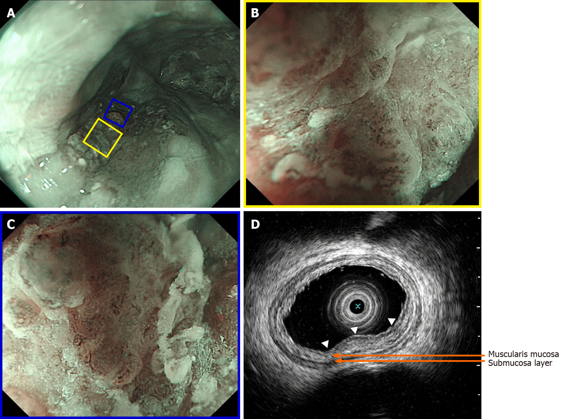
Figure 2 The lesion upon narrow-band imaging.
A: The lesion upon narrow-band imaging; B: Magnified view with magnifying endoscopy with narrow-band imaging of the yellow square in (A) revealing that under the membranous substance and mucous there were intrapapillary capillary loops with morphological irregularity, classified as type B1; C: Magnified view with magnifying endoscopy with narrow-band imaging of the blue square in (A), revealing that in the slightly elevated central part of lesion, irregularly and dendritically branched abnormal vessels without loop formation were observed, classified as type B2; D: Endoscopic ultrasonography revealing a hypoechoic mass (white arrows) confined to the mucosa layer, which did not seem to disrupt the muscularis mucosa completely, and the submucosa layer was intact.
- Citation: Liu GY, Zhang JX, Rong L, Nian WD, Nian BX, Tian Y. Esophageal superficial adenosquamous carcinoma resected by endoscopic submucosal dissection: A rare case report. World J Clin Cases 2021; 9(6): 1336-1342
- URL: https://www.wjgnet.com/2307-8960/full/v9/i6/1336.htm
- DOI: https://dx.doi.org/10.12998/wjcc.v9.i6.1336


