Copyright
©The Author(s) 2021.
World J Clin Cases. Feb 16, 2021; 9(5): 1168-1174
Published online Feb 16, 2021. doi: 10.12998/wjcc.v9.i5.1168
Published online Feb 16, 2021. doi: 10.12998/wjcc.v9.i5.1168
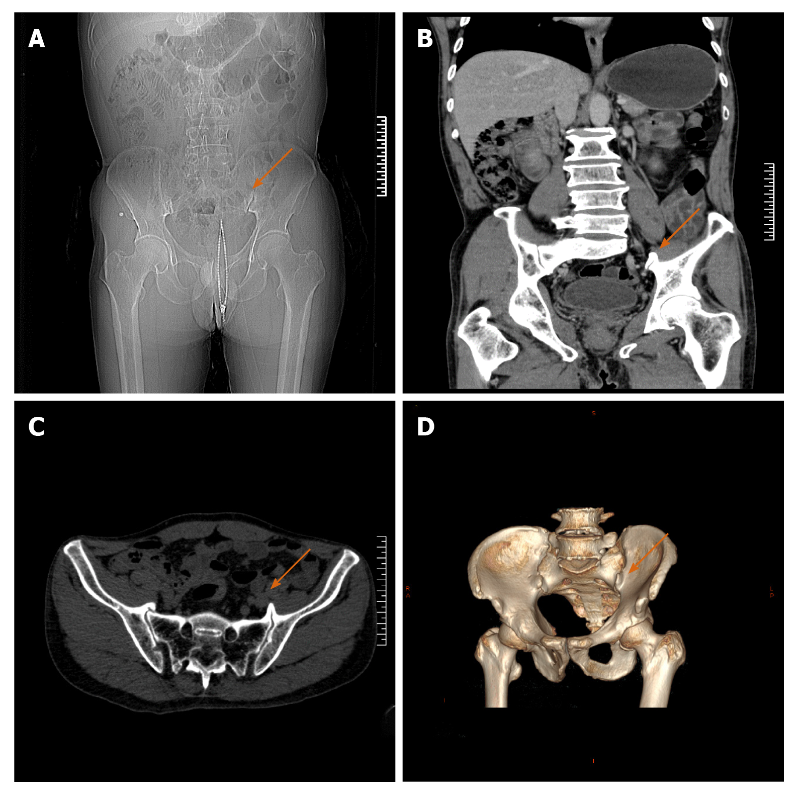
Figure 2 Pelvic computed tomography scan plus 3D reconstruction before the operation.
A: Anteroposterior radiograph of the pelvis; B: Coronal computed tomography scan showing the osteophyte in the left sacroiliac joint, as indicated by the orange arrow; C: Horizontal scan showing that the left osteophyte was more prominent; D: 3D reconstruction revealing the osteophytes that were in the anterior lower part of both sacroiliac joints, with the left osteophyte being larger, as indicated by the orange arrow in the figure.
- Citation: Cai MD, Zhang HF, Fan YG, Su XJ, Xia L. Obturator nerve impingement caused by an osteophyte in the sacroiliac joint: A case report. World J Clin Cases 2021; 9(5): 1168-1174
- URL: https://www.wjgnet.com/2307-8960/full/v9/i5/1168.htm
- DOI: https://dx.doi.org/10.12998/wjcc.v9.i5.1168


