Copyright
©The Author(s) 2021.
World J Clin Cases. Feb 16, 2021; 9(5): 1168-1174
Published online Feb 16, 2021. doi: 10.12998/wjcc.v9.i5.1168
Published online Feb 16, 2021. doi: 10.12998/wjcc.v9.i5.1168
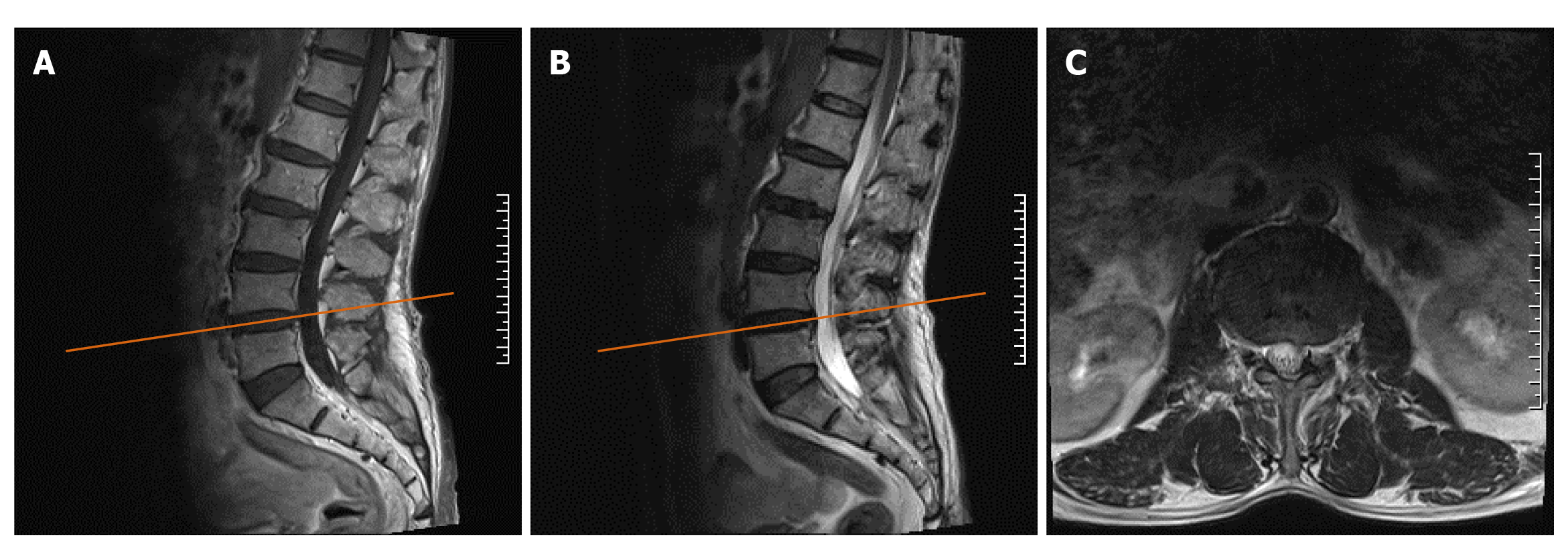
Figure 1 Plain magnetic resonance imaging of the lumbar before the operation.
A: T1 sagittal image showing intervertebral disc bulging at the L4/5 level, which caused slight compression of the anterior edge of the dural sac but not to the extent that causes neurological symptoms; B: T2 sagittal image of the L4/5 level; C: The horizontal image correlated with the L4/5 intervertebral space, as indicated by the yellow scout line in the panel.
- Citation: Cai MD, Zhang HF, Fan YG, Su XJ, Xia L. Obturator nerve impingement caused by an osteophyte in the sacroiliac joint: A case report. World J Clin Cases 2021; 9(5): 1168-1174
- URL: https://www.wjgnet.com/2307-8960/full/v9/i5/1168.htm
- DOI: https://dx.doi.org/10.12998/wjcc.v9.i5.1168


