Copyright
©The Author(s) 2021.
World J Clin Cases. Feb 6, 2021; 9(4): 960-969
Published online Feb 6, 2021. doi: 10.12998/wjcc.v9.i4.960
Published online Feb 6, 2021. doi: 10.12998/wjcc.v9.i4.960
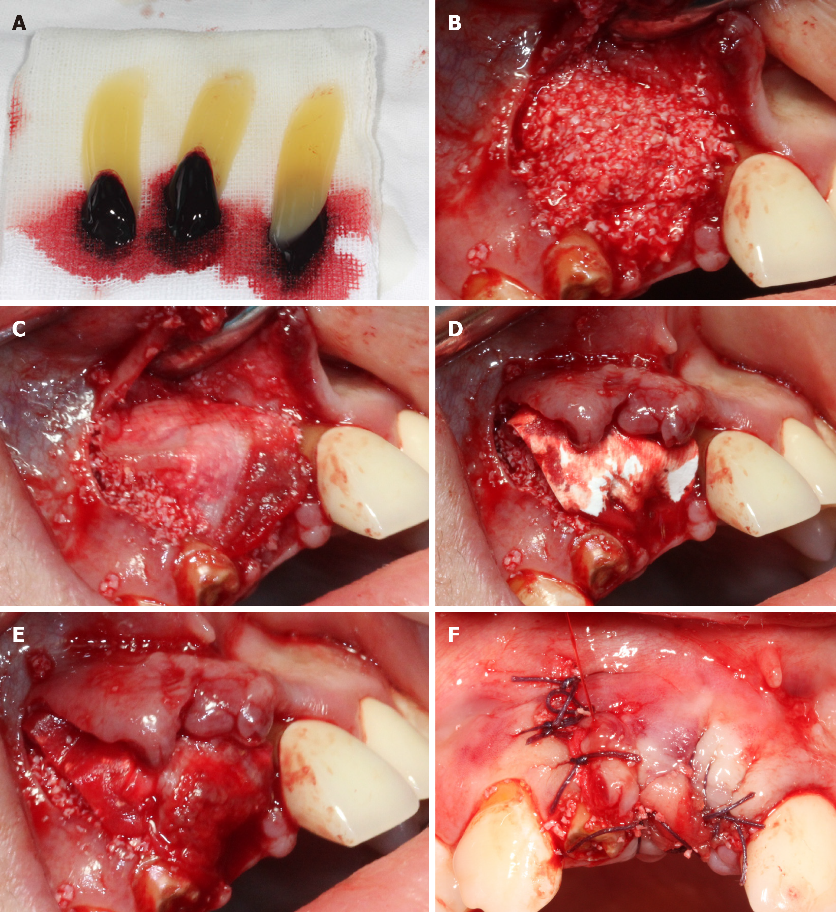
Figure 3 Representational images and illustrations describing each step of covering the barrier membrane.
A: Platelet-rich fibrin (PRF) clots were obtained into the test tube through whole blood centrifuged immediately at 3000 rpm for 10 min; B: PRF clot was mixed with 0.25 g bone substitute (Bio-Oss) then placed on the implant and the bone defect; C: A piece of PRF clot was pressed into a membrane by sterile gauze and placed on the surface of Bio-Oss-PRF mixture; D: Placement of collagen membrane; E: The other PRF membrane was placed on the surface of the collagen membrane; F: Tension-free suture.
- Citation: Fang J, Xin XR, Li W, Wang HC, Lv HX, Zhou YM. Immediate implant placement in combination with platelet rich-fibrin into extraction sites with periapical infection in the esthetic zone: A case report and review of literature. World J Clin Cases 2021; 9(4): 960-969
- URL: https://www.wjgnet.com/2307-8960/full/v9/i4/960.htm
- DOI: https://dx.doi.org/10.12998/wjcc.v9.i4.960


