Copyright
©The Author(s) 2021.
World J Clin Cases. Feb 6, 2021; 9(4): 878-885
Published online Feb 6, 2021. doi: 10.12998/wjcc.v9.i4.878
Published online Feb 6, 2021. doi: 10.12998/wjcc.v9.i4.878
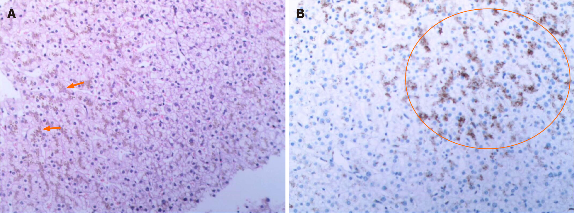
Figure 2 Imaging results.
A: Hematoxylin and eosin staining of liver cells showing a large number of pigment granule deposits (orange arrow) in a patient with suspected Dubin-Johnson syndrome (DJS); original magnification × 20; B: Hepatic tissue immunohistochemistry results showing that liquified or vacuolized liver cells (orange circle), which indicates DJS.
- Citation: Wu H, Zhao XK, Zhu JJ. Clinical characteristics and ABCC2 genotype in Dubin-Johnson syndrome: A case report and review of the literature. World J Clin Cases 2021; 9(4): 878-885
- URL: https://www.wjgnet.com/2307-8960/full/v9/i4/878.htm
- DOI: https://dx.doi.org/10.12998/wjcc.v9.i4.878


