
















©The Author(s) 2021.
World J Clin Cases. Feb 6, 2021; 9(4): 871-877
Published online Feb 6, 2021. doi: 10.12998/wjcc.v9.i4.871
Published online Feb 6, 2021. doi: 10.12998/wjcc.v9.i4.871
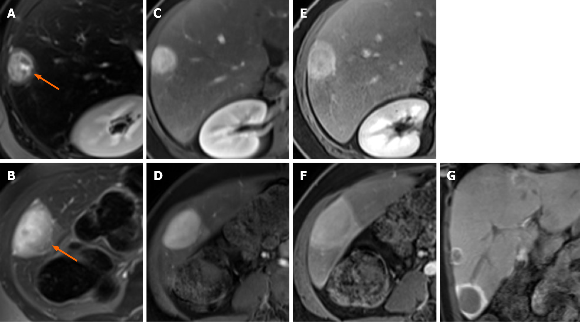
Figure 2 A 17-year-old man glycogen storage disease type I undergoing gadoxetate disodium–enhanced magnetic resonance imaging exam.
A-D: On (A and B) T2-weighted images, two peripheral lesions (arrows) show a markedly heterogenous hyperintense core compared to the surrounding liver and a slightly hyperintense rim. T1-weighted three-dimensional spoiled gradient-echo sequences demonstrate enhancement of the core of the lesions during the arterial phase (C and D); E and F: During the portal venous phase, enhancement persists in the core of the lesions, whilst enhancement appears at the periphery of the lesions; G: During the hepatobiliary phase, the lesions show hypointensity of the central core compared with the background liver and marked contrast retention at the periphery of the lesions.
- Citation: Vernuccio F, Austin S, Meyer M, Guy CD, Kishnani PS, Marin D. "Bull’s eye” appearance of hepatocellular adenomas in patients with glycogen storage disease type I — atypical magnetic resonance imaging findings: Two case reports. World J Clin Cases 2021; 9(4): 871-877
- URL: https://www.wjgnet.com/2307-8960/full/v9/i4/871.htm
- DOI: https://dx.doi.org/10.12998/wjcc.v9.i4.871