Copyright
©The Author(s) 2021.
World J Clin Cases. Dec 26, 2021; 9(36): 11448-11456
Published online Dec 26, 2021. doi: 10.12998/wjcc.v9.i36.11448
Published online Dec 26, 2021. doi: 10.12998/wjcc.v9.i36.11448
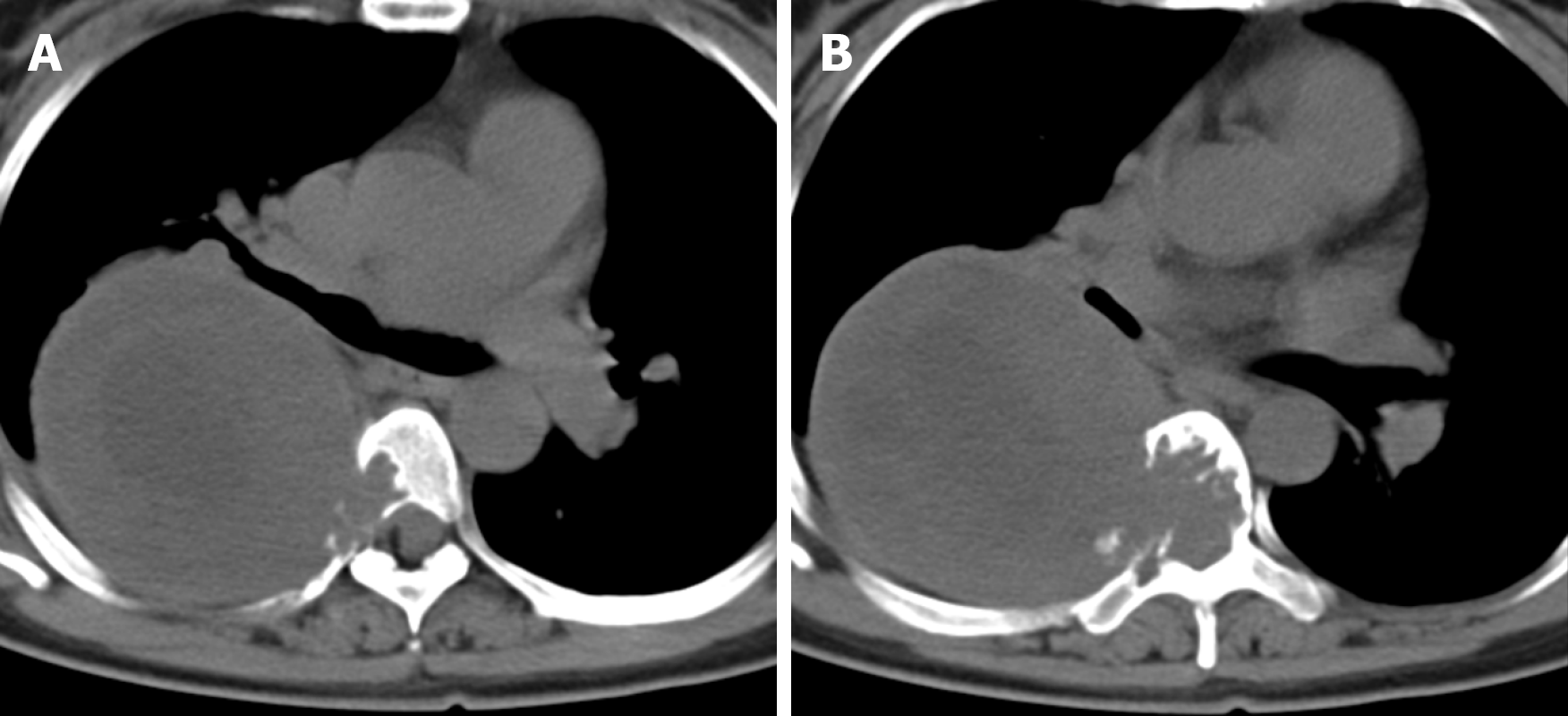
Figure 1 Axial computed tomography revealed bone destruction visible in the T5 (A) and T6 (B) vertebrae and the right accessory and part of the right rib, and irregular soft tissue density shadows were seen in the T5 and T6 vertebrae, which protruded into the thoracic cavity to the right.
- Citation: Zhou Y, Liu CZ, Zhang SY, Wang HY, Nath Varma S, Cao LQ, Hou TT, Li X, Yao BJ. Giant schwannoma of thoracic vertebra: A case report. World J Clin Cases 2021; 9(36): 11448-11456
- URL: https://www.wjgnet.com/2307-8960/full/v9/i36/11448.htm
- DOI: https://dx.doi.org/10.12998/wjcc.v9.i36.11448


