
















©The Author(s) 2021.
World J Clin Cases. Dec 26, 2021; 9(36): 11400-11405
Published online Dec 26, 2021. doi: 10.12998/wjcc.v9.i36.11400
Published online Dec 26, 2021. doi: 10.12998/wjcc.v9.i36.11400
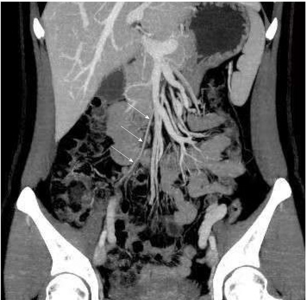
Figure 2 Follow-up computed tomography scan after 1 mo.
Coronal reformatted image with maximum intensity projection showed the ileocolic artery (arrow). The site of previous thrombophlebitis in the ileocolic vein had disappeared, which indicated that the vein was completely occluded and that collateral circulation was established.
- Citation: Yang F, Guo XC, Rao XL, Sun L, Xu L. Acute appendicitis complicated by mesenteric vein thrombosis: A case report. World J Clin Cases 2021; 9(36): 11400-11405
- URL: https://www.wjgnet.com/2307-8960/full/v9/i36/11400.htm
- DOI: https://dx.doi.org/10.12998/wjcc.v9.i36.11400