
















©The Author(s) 2021.
World J Clin Cases. Dec 6, 2021; 9(34): 10723-10727
Published online Dec 6, 2021. doi: 10.12998/wjcc.v9.i34.10723
Published online Dec 6, 2021. doi: 10.12998/wjcc.v9.i34.10723
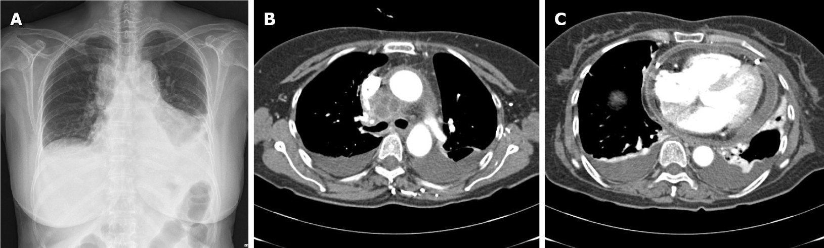
Figure 2 Chest X-ray radiograph and computed tomography.
A: Chest X-ray radiograph showed both pleural effusion and cardiomegaly; B and C: Chest computed tomography performed 7 d after ultrasound-guided transbronchial needle aspiration demonstrated (B) an increased size and mediastinal abscess formation in the right paratracheal area and (C) newly developed moderate amounts of pericardial effusion with diffuse pericardial thickening.
- Citation: Koh JS, Kim YJ, Kang DH, Lee JE, Lee SI. Severe mediastinitis and pericarditis after endobronchial ultrasound-guided transbronchial needle aspiration: A case report. World J Clin Cases 2021; 9(34): 10723-10727
- URL: https://www.wjgnet.com/2307-8960/full/v9/i34/10723.htm
- DOI: https://dx.doi.org/10.12998/wjcc.v9.i34.10723