Copyright
©The Author(s) 2021.
World J Clin Cases. Dec 6, 2021; 9(34): 10715-10722
Published online Dec 6, 2021. doi: 10.12998/wjcc.v9.i34.10715
Published online Dec 6, 2021. doi: 10.12998/wjcc.v9.i34.10715
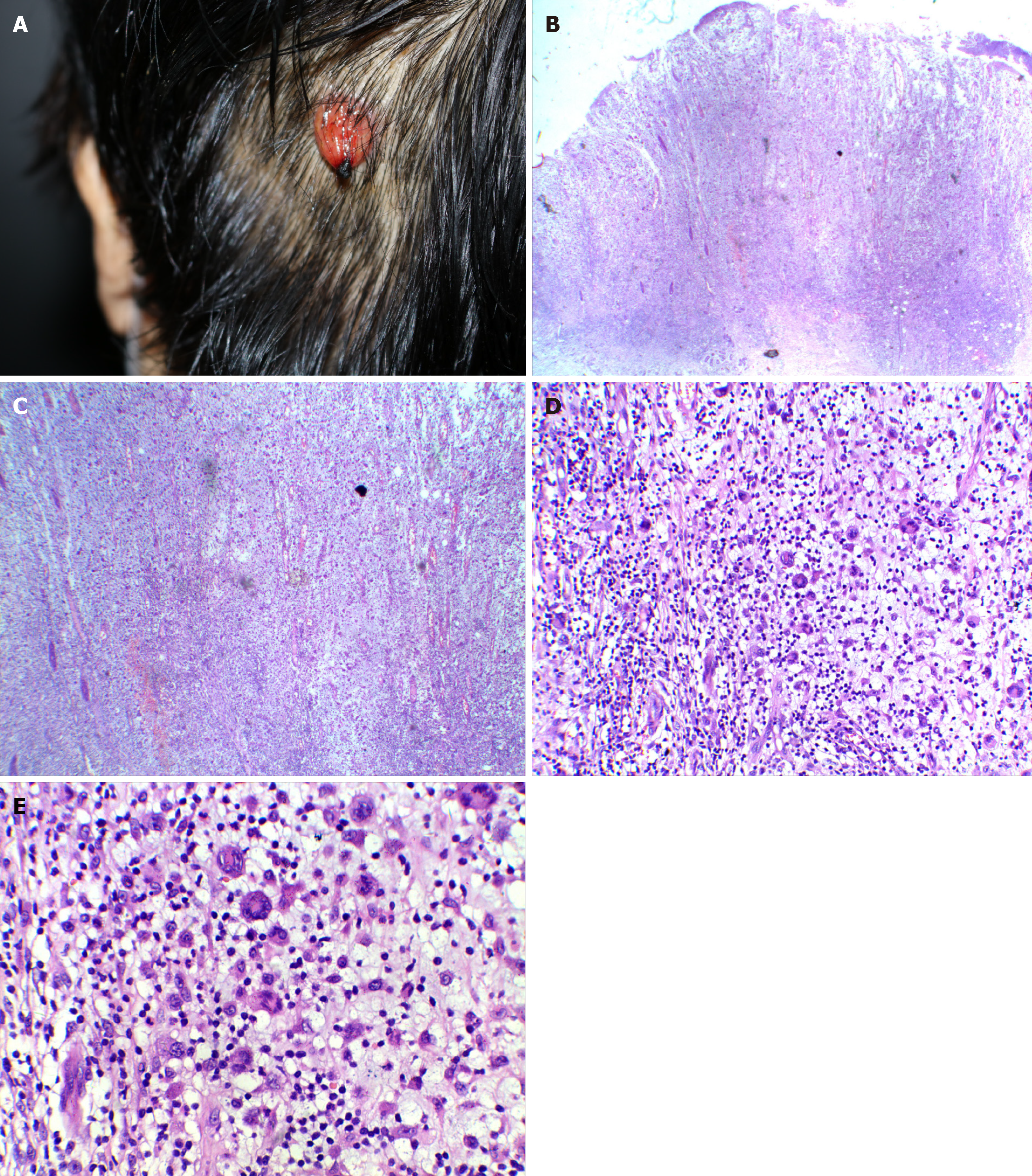
Figure 1 Morphology and skin biopsy of the scalp lesion.
The patient was admitted for the fourth time and was diagnosed as chronic lymphocytic leukemia/small lymphocytic lymphoma complicated with skin Langerhans cell sarcoma). A: Lesion on the left side of the head, looking like a hanging bag, with a soft texture, a slightly hard base, and ulceration and blood scabs at the tip; B: Skin tissue local ulcer formation (hematoxylin and eosin [HE], × 10); C: Ulcer showed mixed cell proliferation, vascular proliferation (HE, × 20); D: Some cells were enlarged, with a rich cytoplasm, light staining, mononuclear, binuclear, multinucleated, or lobulated nucleus; phagocytosis of lymphocytes could be seen in some cytoplasm. In the background, there were more scattered lymphocytes, neutrophils, and local interstitial mucoid degeneration pustular folliculitis (HE, × 100); E: Some cells were enlarged, with a rich cytoplasm, light staining, mononuclear, binuclear, multinucleated, or lobulated nucleus; phagocytosis of lymphocytes could be seen in some cytoplasm. In the background, there were more scattered lymphocytes, neutrophils, and local interstitial mucoid degeneration pustular folliculitis (HE, × 200).
- Citation: Li SY, Wang Y, Wang LH. Chronic lymphocytic leukemia/small lymphocytic lymphoma complicated with skin Langerhans cell sarcoma: A case report . World J Clin Cases 2021; 9(34): 10715-10722
- URL: https://www.wjgnet.com/2307-8960/full/v9/i34/10715.htm
- DOI: https://dx.doi.org/10.12998/wjcc.v9.i34.10715


