Copyright
©The Author(s) 2021.
World J Clin Cases. Dec 6, 2021; 9(34): 10708-10714
Published online Dec 6, 2021. doi: 10.12998/wjcc.v9.i34.10708
Published online Dec 6, 2021. doi: 10.12998/wjcc.v9.i34.10708
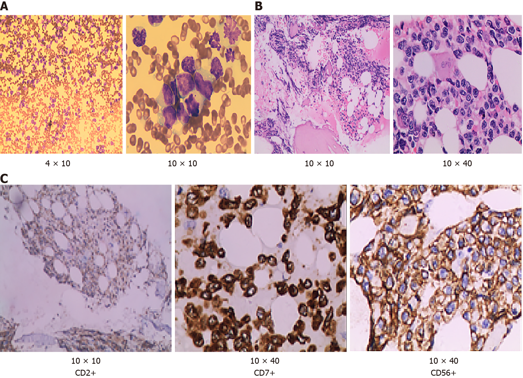
Figure 2 Hematoxylin–eosin staining.
A: Bone marrow cell morphology test showed that cells with unknown classification and abnormality were easy to see, accounting for 94% (magnification: 4 × 10 and 10 × 10); B: Bone marrow biopsy test showed that hematopoietic tissue proliferation was heterogeneous, adipose tissue hyperplasia was decreased, granulocyte and erythrocytic proliferation was decreased, megakaryocytic hyperplasia (0–4/high-power field) was scattered (magnification: 10 × 10 and 10 × 40); C: Immunohistochemical of bone marrow biopsy showed that the atypical cells were positive for CD2, cCD3, CD7, CD20, CD34, CD68, CD56, and negative for sCD3 (magnification: 10 × 10; 10 × 40 and 10 × 10).
- Citation: Peng XH, Zhang LS, Li LJ, Guo XJ, Liu Y. Aggressive natural killer cell leukemia with skin manifestation associated with hemophagocytic lymphohistiocytosis: A case report. World J Clin Cases 2021; 9(34): 10708-10714
- URL: https://www.wjgnet.com/2307-8960/full/v9/i34/10708.htm
- DOI: https://dx.doi.org/10.12998/wjcc.v9.i34.10708


