Copyright
©The Author(s) 2021.
World J Clin Cases. Dec 6, 2021; 9(34): 10696-10701
Published online Dec 6, 2021. doi: 10.12998/wjcc.v9.i34.10696
Published online Dec 6, 2021. doi: 10.12998/wjcc.v9.i34.10696
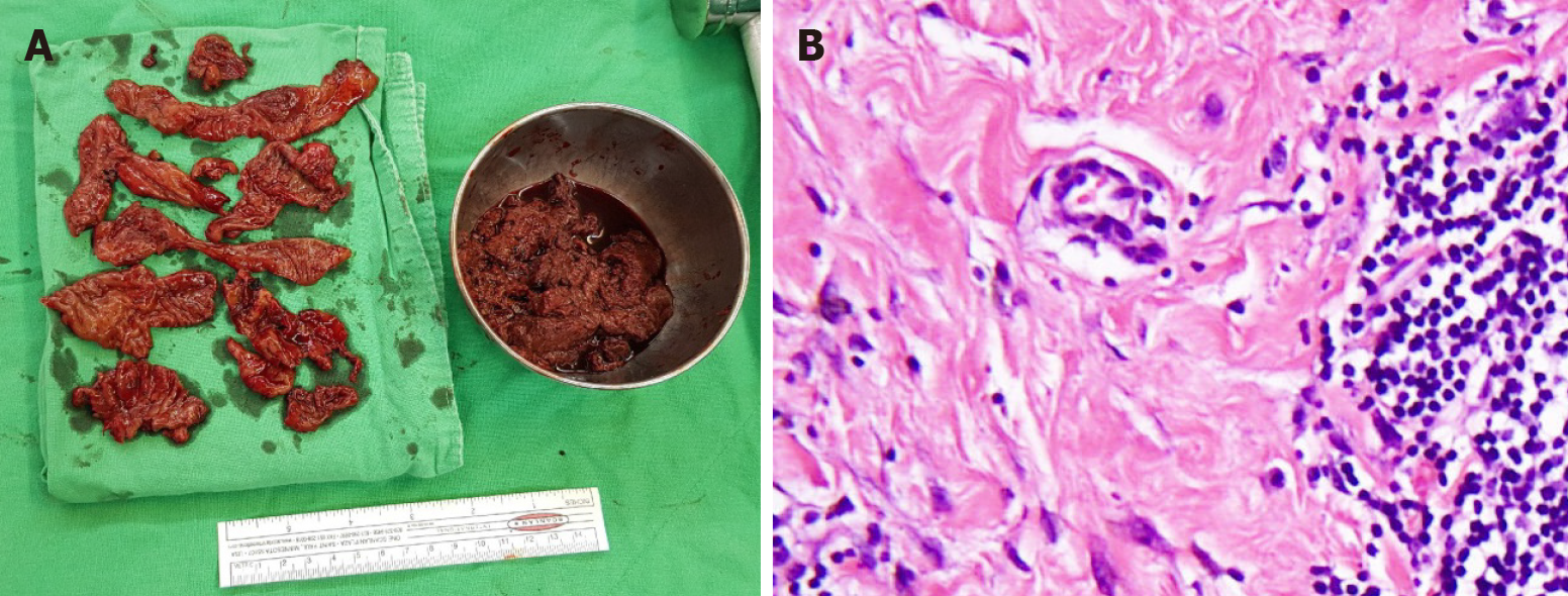
Figure 3 Gross photo and pathological section of resected pseudotumor.
A: Photograph of debrided necrotic tissues taken from arthrotomy and removal of pseudotumor; B: Synovial lining cell hyperplasia with lymphocytic cells infiltration and Stromal fibroplasia, with the histological diagnosis of aseptic aseptic lymphocyte-dominant vasculitis-associated lesion.
- Citation: Lin IH, Tsai CH. Tigecycline sclerotherapy for recurrent pseudotumor in aseptic lymphocyte-dominant vasculitis-associated lesion after metal-on-metal total hip arthroplasty: A case report. World J Clin Cases 2021; 9(34): 10696-10701
- URL: https://www.wjgnet.com/2307-8960/full/v9/i34/10696.htm
- DOI: https://dx.doi.org/10.12998/wjcc.v9.i34.10696


