Copyright
©The Author(s) 2021.
World J Clin Cases. Dec 6, 2021; 9(34): 10681-10688
Published online Dec 6, 2021. doi: 10.12998/wjcc.v9.i34.10681
Published online Dec 6, 2021. doi: 10.12998/wjcc.v9.i34.10681
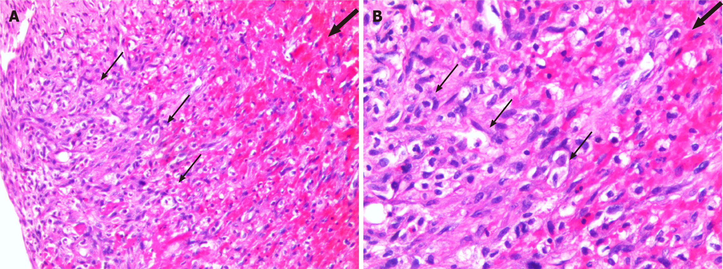
Figure 3 Histological features of the epidural mass.
A: Hematoxylin-eosin (HE); × 100; B: HE × 200. Histopathological pictomicrograph shows dilated thin-walled vessels lined by a monolayer of obese endothelial cells (thin arrows). The lumen appears to be filled with organizing thrombi (thick arrow).
- Citation: Gu HL, Zheng XQ, Zhan SQ, Chang YB. Intravascular papillary endothelial hyperplasia as a rare cause of cervicothoracic spinal cord compression: A case report. World J Clin Cases 2021; 9(34): 10681-10688
- URL: https://www.wjgnet.com/2307-8960/full/v9/i34/10681.htm
- DOI: https://dx.doi.org/10.12998/wjcc.v9.i34.10681


