Copyright
©The Author(s) 2021.
World J Clin Cases. Dec 6, 2021; 9(34): 10518-10529
Published online Dec 6, 2021. doi: 10.12998/wjcc.v9.i34.10518
Published online Dec 6, 2021. doi: 10.12998/wjcc.v9.i34.10518
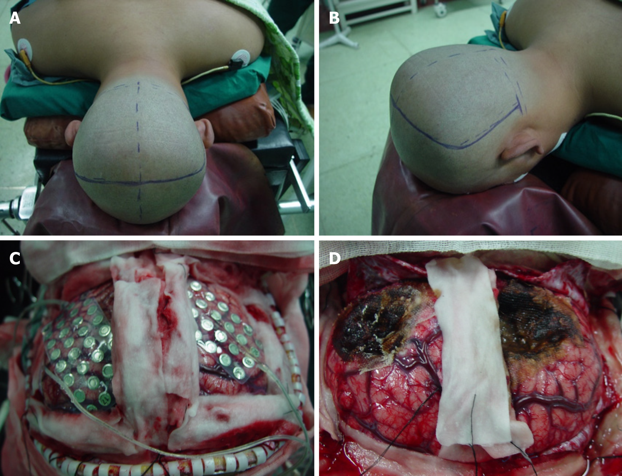
Figure 1 Surgical resection of bilateral occipital lesions.
A and B: The extent of the bilateral occipital craniotomy; C: Intracranial cortical electrodes were placed on the surface of the bilateral occipital lobe during surgery to enable monitoring of the electroencephalography; D: Photograph taken after resection of the lesions in the bilateral occipital lobe.
- Citation: Lyu YE, Xu XF, Dai S, Feng M, Shen SP, Zhang GZ, Ju HY, Wang Y, Dong XB, Xu B. Resection of bilateral occipital lobe lesions during a single operation as a treatment for bilateral occipital lobe epilepsy. World J Clin Cases 2021; 9(34): 10518-10529
- URL: https://www.wjgnet.com/2307-8960/full/v9/i34/10518.htm
- DOI: https://dx.doi.org/10.12998/wjcc.v9.i34.10518


