Copyright
©The Author(s) 2021.
World J Clin Cases. Dec 6, 2021; 9(34): 10494-10506
Published online Dec 6, 2021. doi: 10.12998/wjcc.v9.i34.10494
Published online Dec 6, 2021. doi: 10.12998/wjcc.v9.i34.10494
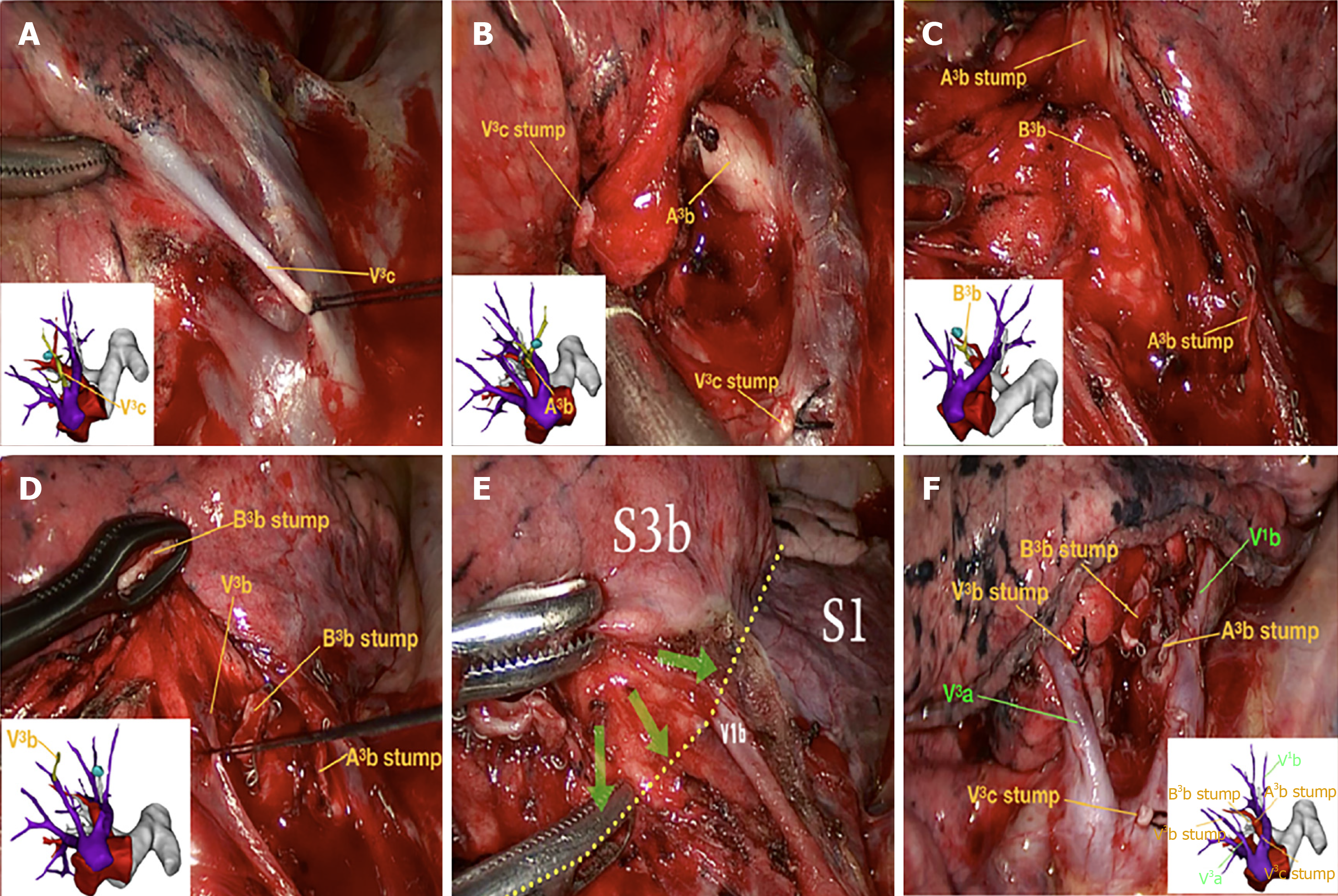
Figure 4 Illustration of the precise surgical procedure navigated by three-dimensional computed-tomography bronchography and angiography.
With the assistance of three-dimensional computed-tomography bronchography and angiography (3D-CTBA), we meticulously performed thoracoscopic segmentectomy of the RS3b. A series of techniques were involved in this procedure, including location of the pulmonary nodules, resection of targeted vessels and bronchi, preservation of intersegmental veins, and identification of the intersegmental demarcation. A–D: Images showing the resection sequence for the targeted vessels and bronchi, including the V3c, A3b, B3b, and V3b, respectively. The intersegmental demarcation (yellow dotted line) was defined by the improved inflation–deflation method, assisted by 3D-CTBA. The intersegmental veins V1b and V3a were carefully preserved. The surgeons precisely identified, separated, and dissected the targeted segment based on the cone-shaped principle; E, F: View of the hilum after RS3b removal reveals stumps of targeted vessels and bronchi, which was completely consistent with the preoperative 3D-CTBA images.
- Citation: Wu YJ, Shi QT, Zhang Y, Wang YL. Thoracoscopic segmentectomy and lobectomy assisted by three-dimensional computed-tomography bronchography and angiography for the treatment of primary lung cancer. World J Clin Cases 2021; 9(34): 10494-10506
- URL: https://www.wjgnet.com/2307-8960/full/v9/i34/10494.htm
- DOI: https://dx.doi.org/10.12998/wjcc.v9.i34.10494


