Copyright
©The Author(s) 2021.
World J Clin Cases. Dec 6, 2021; 9(34): 10494-10506
Published online Dec 6, 2021. doi: 10.12998/wjcc.v9.i34.10494
Published online Dec 6, 2021. doi: 10.12998/wjcc.v9.i34.10494
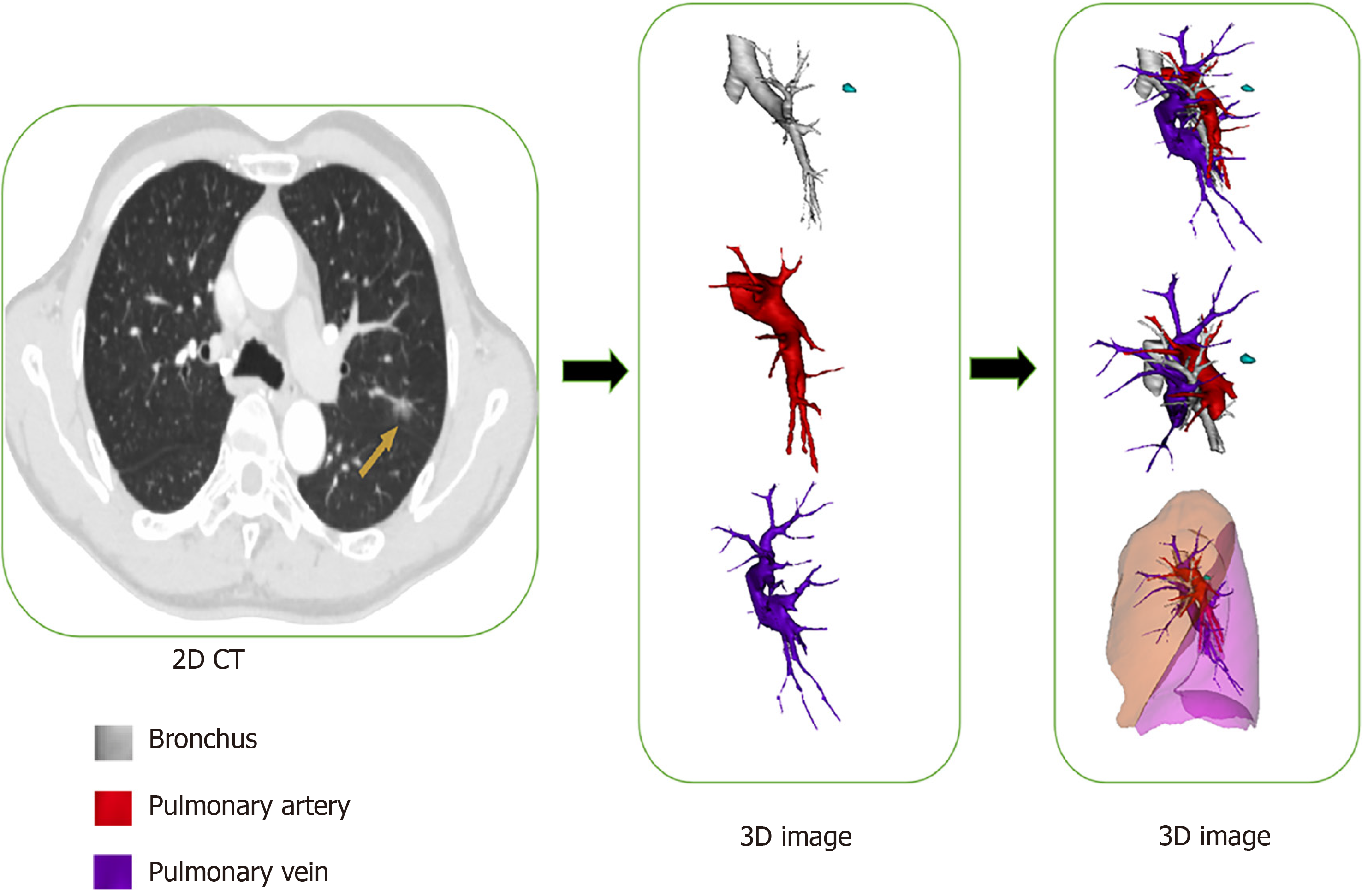
Figure 1 Preoperative flow diagram of three-dimensional reconstruction technique using Mimics software.
The pulmonary bronchus, artery and vein were distinguished from one another and marked out with different colors: white, bronchus; red, pulmonary artery; purple, pulmonary vein. 3D: 3-dimensional; CT: Computed-tomography.
- Citation: Wu YJ, Shi QT, Zhang Y, Wang YL. Thoracoscopic segmentectomy and lobectomy assisted by three-dimensional computed-tomography bronchography and angiography for the treatment of primary lung cancer. World J Clin Cases 2021; 9(34): 10494-10506
- URL: https://www.wjgnet.com/2307-8960/full/v9/i34/10494.htm
- DOI: https://dx.doi.org/10.12998/wjcc.v9.i34.10494


