Copyright
©The Author(s) 2021.
World J Clin Cases. Nov 16, 2021; 9(32): 9889-9895
Published online Nov 16, 2021. doi: 10.12998/wjcc.v9.i32.9889
Published online Nov 16, 2021. doi: 10.12998/wjcc.v9.i32.9889
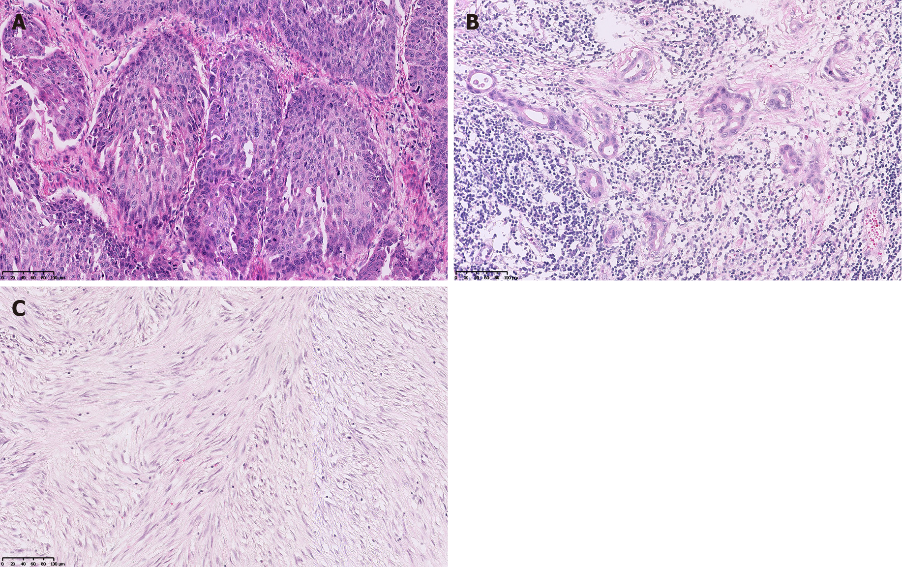
Figure 3 Postoperative pathology.
A: Postoperative pathology showed that the esophageal tumor was a moderately differentiated squamous cell carcinoma with lymph nodes metastases (pT3N2M0, G2, stage IIIB); B: Postoperative pathology showed that the gastric tumor was a moderately to poorly differentiated adenocarcinoma (tubular adenocarcinoma and signet-ring cell carcinomaa, Laurén mixed type) with lymph node metastases (pT3N2M0, G2-G3, stage IIIA); C: Postoperative pathology showed that the jejunal tumor was a gastrointestinal stromal tumor of spindle cell type (high-risk).
- Citation: Li Y, Ye LS, Hu B. Synchronous multiple primary malignancies of the esophagus, stomach, and jejunum: A case report. World J Clin Cases 2021; 9(32): 9889-9895
- URL: https://www.wjgnet.com/2307-8960/full/v9/i32/9889.htm
- DOI: https://dx.doi.org/10.12998/wjcc.v9.i32.9889


