Copyright
©The Author(s) 2021.
World J Clin Cases. Nov 6, 2021; 9(31): 9652-9661
Published online Nov 6, 2021. doi: 10.12998/wjcc.v9.i31.9652
Published online Nov 6, 2021. doi: 10.12998/wjcc.v9.i31.9652
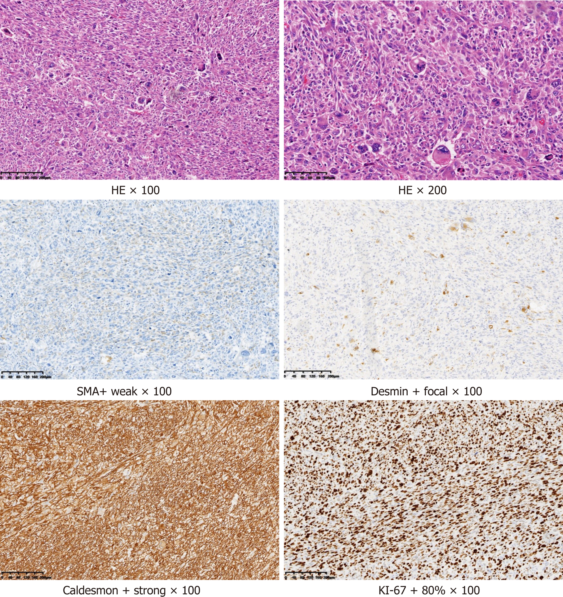
Figure 4 Postoperative pathological picture.
The size of the resected tumor tissue was about 12 cm × 7 cm × 4.5 cm, with hemorrhage and necrosis. Under the microscope (hematoxylin and eosin staining), the tumor cells were short fusiform or epithelioid with obvious atypia. Multinucleated and pleomorphic giant cells were also present. Mitosis indicating active growth was present. Immunohistochemistry revealed that the tumor was positive for smooth muscle actin, desmin, and caldesmon, suggesting a smooth muscle origin, and was negative for cytokeratin, epithelial membrane antigen, synaptophysin, S-100, chromogranin A, and cluster of differentiation 56 (CD56), suggesting that the tumor was not epithelial, neurogenic, neuroendocrine, or neuroectodermal. Ki-67, an antigen index of cell proliferation, had a value of 60%. The higher the index, the higher the risk of malignancy. Cytokeratin, epithelial membrane antigen, synaptophysin, S-100, chromogranin A, and CD56 were all negative in this case (data not shown), suggesting that the tumor was not epithelial, neurogenic, neuroendocrine, or neuroectodermal. HE: Hematoxylin and eosin.
- Citation: Xie XJ, Jiang TA, Zhao QY. Diagnostic value of contrast-enhanced ultrasonography in mediastinal leiomyosarcoma mimicking aortic hematoma: A case report and review of literature . World J Clin Cases 2021; 9(31): 9652-9661
- URL: https://www.wjgnet.com/2307-8960/full/v9/i31/9652.htm
- DOI: https://dx.doi.org/10.12998/wjcc.v9.i31.9652


