
















©The Author(s) 2021.
World J Clin Cases. Nov 6, 2021; 9(31): 9652-9661
Published online Nov 6, 2021. doi: 10.12998/wjcc.v9.i31.9652
Published online Nov 6, 2021. doi: 10.12998/wjcc.v9.i31.9652
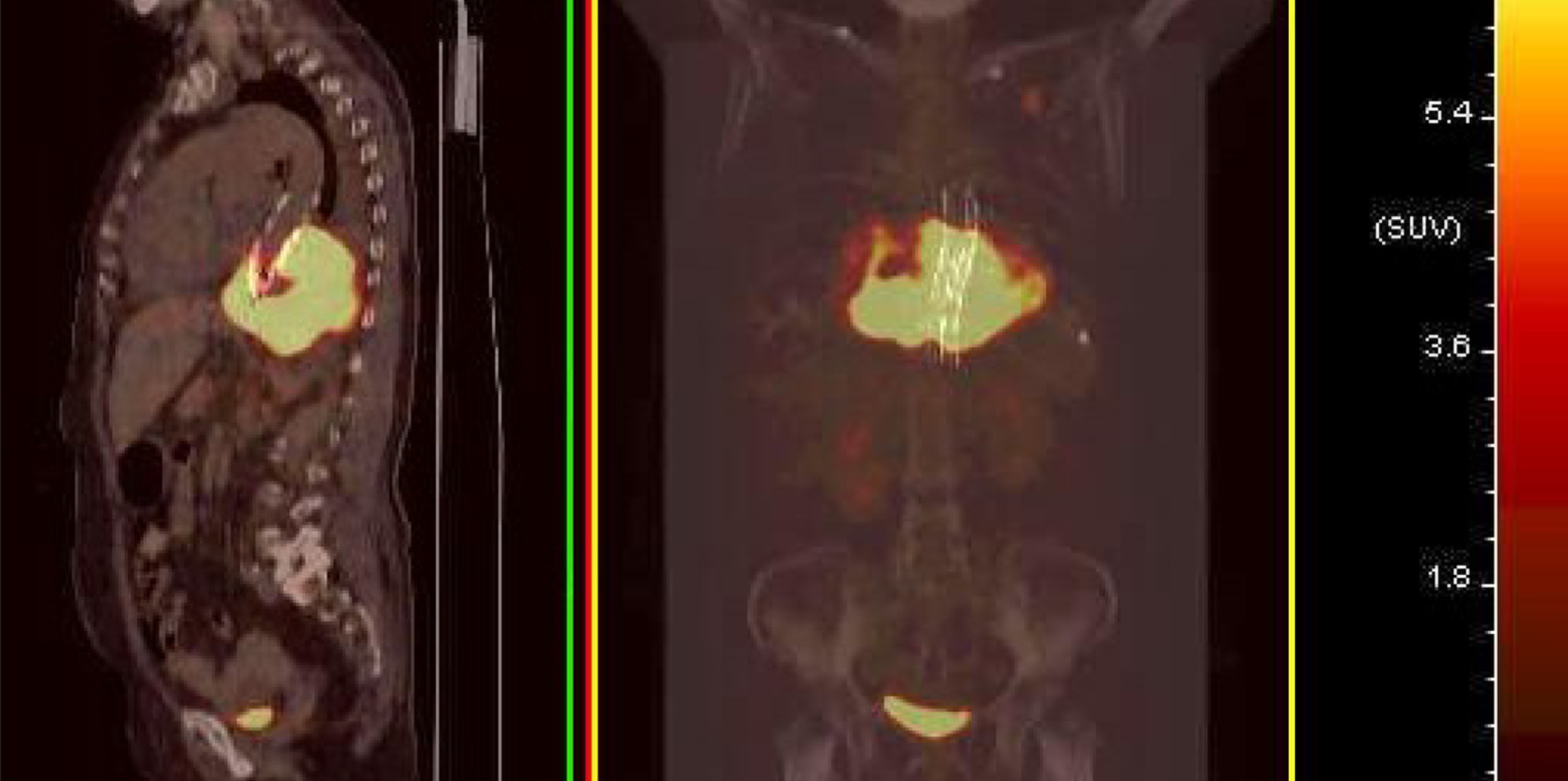
Figure 3 Median sagittal and coronal section images of positron emission tomography/computed tomography.
Positron emission tomography/computed tomography showed that the mass with high fluorodeoxyglucose (FDG) metabolism surrounded the descending aorta. The maximum standardized uptake value (SUV) was 28.8. A nodule with a high FDG metabolism was observed in the left upper lobe, with a maximum SUV of 9.5. The high metabolic signal in the bladder should represent the excreted drugs. No abnormal increase in FDG metabolism was observed in the rest of the body.
- Citation: Xie XJ, Jiang TA, Zhao QY. Diagnostic value of contrast-enhanced ultrasonography in mediastinal leiomyosarcoma mimicking aortic hematoma: A case report and review of literature . World J Clin Cases 2021; 9(31): 9652-9661
- URL: https://www.wjgnet.com/2307-8960/full/v9/i31/9652.htm
- DOI: https://dx.doi.org/10.12998/wjcc.v9.i31.9652