Copyright
©The Author(s) 2021.
World J Clin Cases. Nov 6, 2021; 9(31): 9623-9628
Published online Nov 6, 2021. doi: 10.12998/wjcc.v9.i31.9623
Published online Nov 6, 2021. doi: 10.12998/wjcc.v9.i31.9623
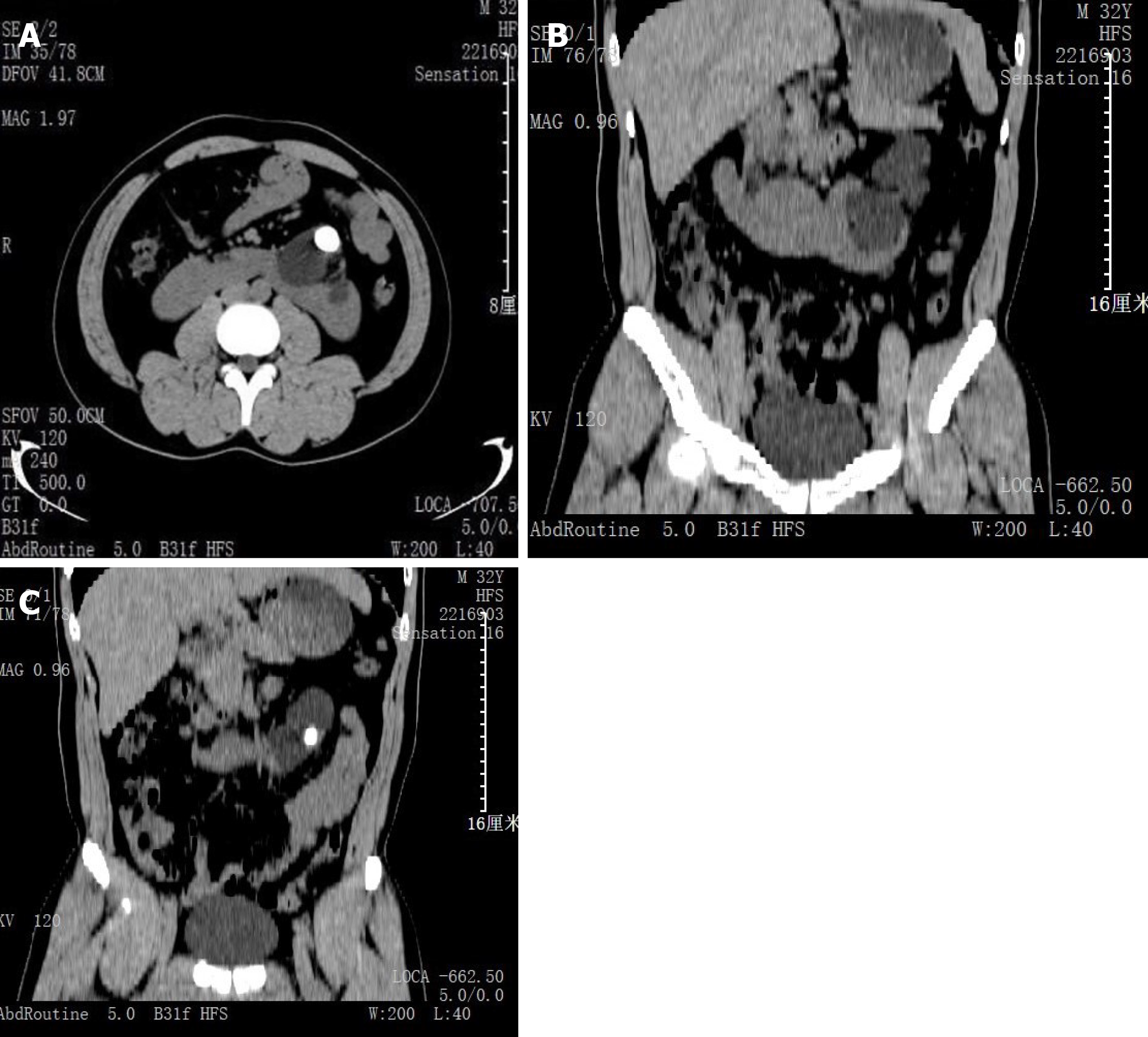
Figure 1 CT images.
A: CT revealed a horseshoe kidney with left renal pelvis calculi; B: Coronal CT slide displayed the lower pole fusion of horseshoe kidney; C: Coronal CT slide displayed the lower pole fusion and location of the stone in the pelvis. CT: Computed tomography.
- Citation: Zhou C, Yan ZJ, Cheng Y, Jiang JH. Bilateral hematoma after tubeless percutaneous nephrolithotomy for unilateral horseshoe kidney stones: A case report. World J Clin Cases 2021; 9(31): 9623-9628
- URL: https://www.wjgnet.com/2307-8960/full/v9/i31/9623.htm
- DOI: https://dx.doi.org/10.12998/wjcc.v9.i31.9623


