
















©The Author(s) 2021.
World J Clin Cases. Nov 6, 2021; 9(31): 9557-9563
Published online Nov 6, 2021. doi: 10.12998/wjcc.v9.i31.9557
Published online Nov 6, 2021. doi: 10.12998/wjcc.v9.i31.9557
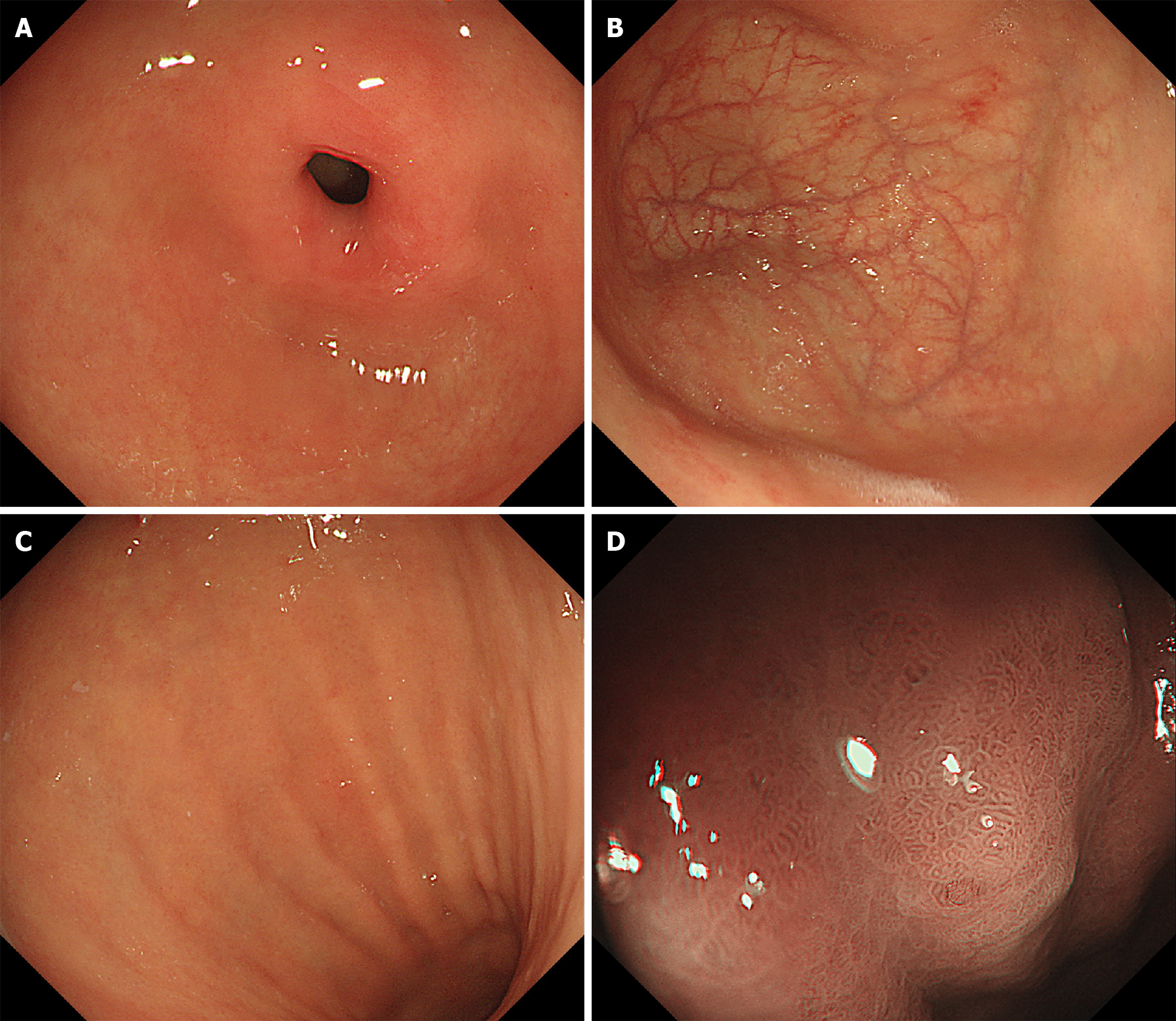
Figure 1 Endoscopic findings.
Endoscopy shows normal mucosa in the gastric antrum (A); and atrophic mucosa in the gastric fundus and body (B and C). Narrow band imaging shows enlarged tubular structures without regular arrangement of collecting venules and pseudopyloric changes in the gastric fundus and body (D).
- Citation: Sun WJ, Ma Q, Liang RZ, Ran YM, Zhang L, Xiao J, Peng YM, Zhan B. Validation of diagnostic strategies of autoimmune atrophic gastritis: A case report. World J Clin Cases 2021; 9(31): 9557-9563
- URL: https://www.wjgnet.com/2307-8960/full/v9/i31/9557.htm
- DOI: https://dx.doi.org/10.12998/wjcc.v9.i31.9557