
















©The Author(s) 2021.
World J Clin Cases. Oct 26, 2021; 9(30): 9285-9294
Published online Oct 26, 2021. doi: 10.12998/wjcc.v9.i30.9285
Published online Oct 26, 2021. doi: 10.12998/wjcc.v9.i30.9285
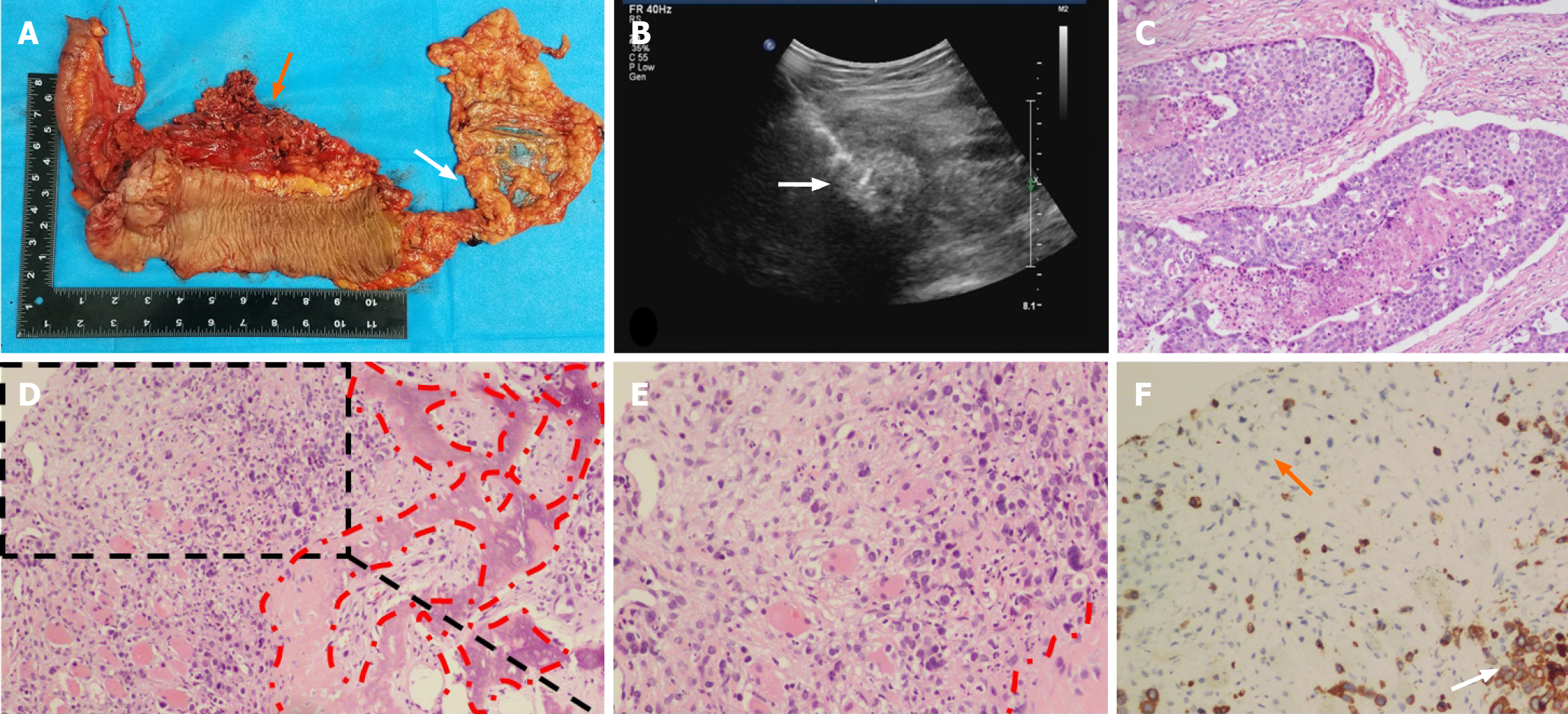
Figure 2 Pathology and immunohistochemistry of the primary lesion and thigh mass.
A: In the resected specimen, the orange arrow points to the tumor location and the white arrow points to the appendix; B: Skeletal muscle metastasis (white arrow) on color ultrasound; C: Hematoxylin-eosin staining of the resected colon cancer specimen (magnification, 40 times); D: H&E staining of an SMM biopsy. The red dashed circle is the bone metaplasia region (magnification, 40 times); E: Part of the enlarged Figure 1D (magnification, 200 times); F: Cytokeratin staining of an SMM biopsy. The orange arrow refers to the fibroblasts in the tumor stroma and the white arrow refers to the metastatic tumor cells (magnification, 200 times).
- Citation: Guo Y, Wang S, Zhao ZY, Li JN, Shang A, Li DL, Wang M. Skeletal muscle metastasis with bone metaplasia from colon cancer: A case report and review of the literature. World J Clin Cases 2021; 9(30): 9285-9294
- URL: https://www.wjgnet.com/2307-8960/full/v9/i30/9285.htm
- DOI: https://dx.doi.org/10.12998/wjcc.v9.i30.9285