
















©The Author(s) 2021.
World J Clin Cases. Oct 26, 2021; 9(30): 9276-9284
Published online Oct 26, 2021. doi: 10.12998/wjcc.v9.i30.9276
Published online Oct 26, 2021. doi: 10.12998/wjcc.v9.i30.9276
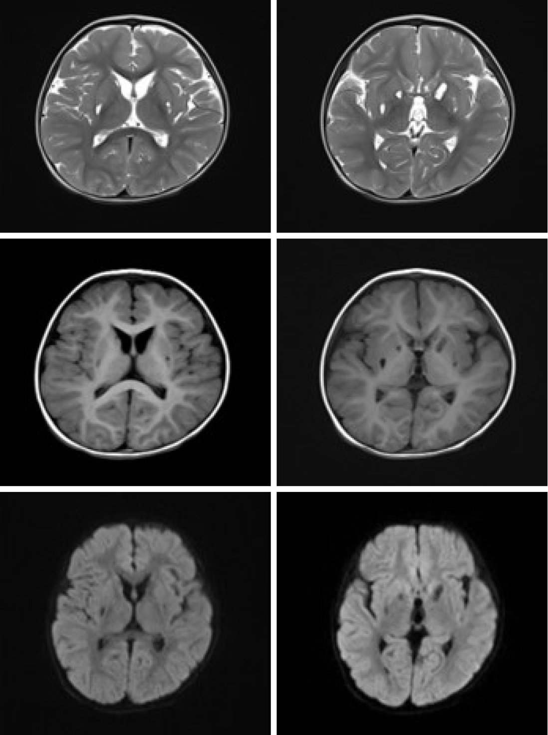
Figure 4 Magnetic resonance images of the patient at 1 year after admission showing that the lesions were completely liquefied and bilateral caudate nuclei were still atrophic.
The liquefaction focus of bilateral lentiform nuclei had no significant improvement.
- Citation: Guo J, Ren D, Guo ZJ, Yu J, Liu F, Zhao RX, Wang Y. Emergence of lesions outside of the basal ganglia and irreversible damage to the basal ganglia with severe β-ketothiolase deficiency: A case report . World J Clin Cases 2021; 9(30): 9276-9284
- URL: https://www.wjgnet.com/2307-8960/full/v9/i30/9276.htm
- DOI: https://dx.doi.org/10.12998/wjcc.v9.i30.9276