Copyright
©The Author(s) 2021.
World J Clin Cases. Oct 26, 2021; 9(30): 9211-9217
Published online Oct 26, 2021. doi: 10.12998/wjcc.v9.i30.9211
Published online Oct 26, 2021. doi: 10.12998/wjcc.v9.i30.9211
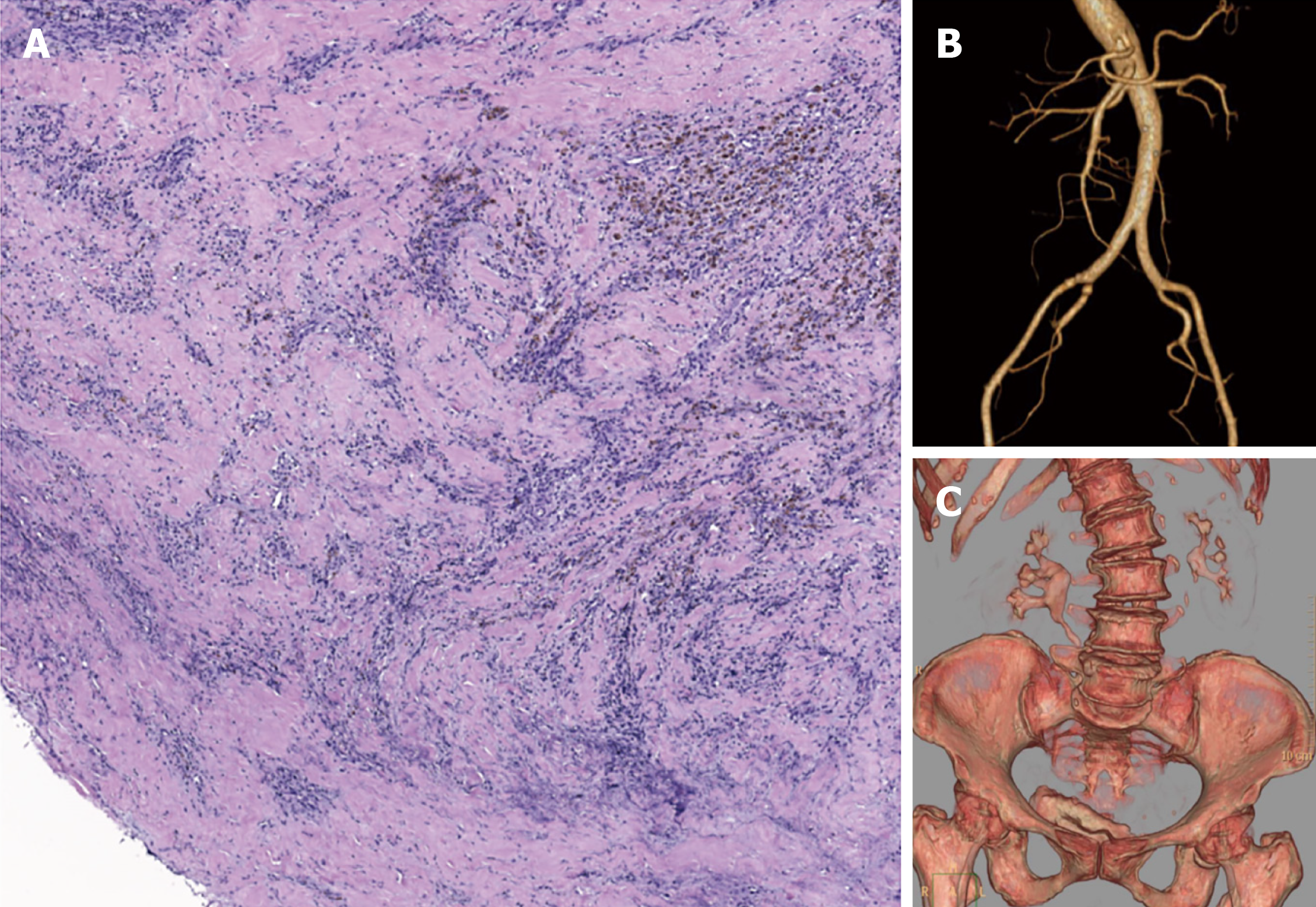
Figure 3 Surgical pathology and 6-mo follow-up computed tomography.
A: Organized thrombus of inferior vena cava with massive inflammatory cell infiltration; B: Computed tomography urography indicated mild right hydronephrosis; C: Computed tomography angiography indicated patency of bilateral iliac arteries.
- Citation: Weng CX, Wang SM, Wang TH, Zhao JC, Yuan D. Successful management of infected right iliac pseudoaneurysm caused by penetration of migrated inferior vena cava filter: A case report. World J Clin Cases 2021; 9(30): 9211-9217
- URL: https://www.wjgnet.com/2307-8960/full/v9/i30/9211.htm
- DOI: https://dx.doi.org/10.12998/wjcc.v9.i30.9211


