Copyright
©The Author(s) 2021.
World J Clin Cases. Oct 26, 2021; 9(30): 9122-9128
Published online Oct 26, 2021. doi: 10.12998/wjcc.v9.i30.9122
Published online Oct 26, 2021. doi: 10.12998/wjcc.v9.i30.9122
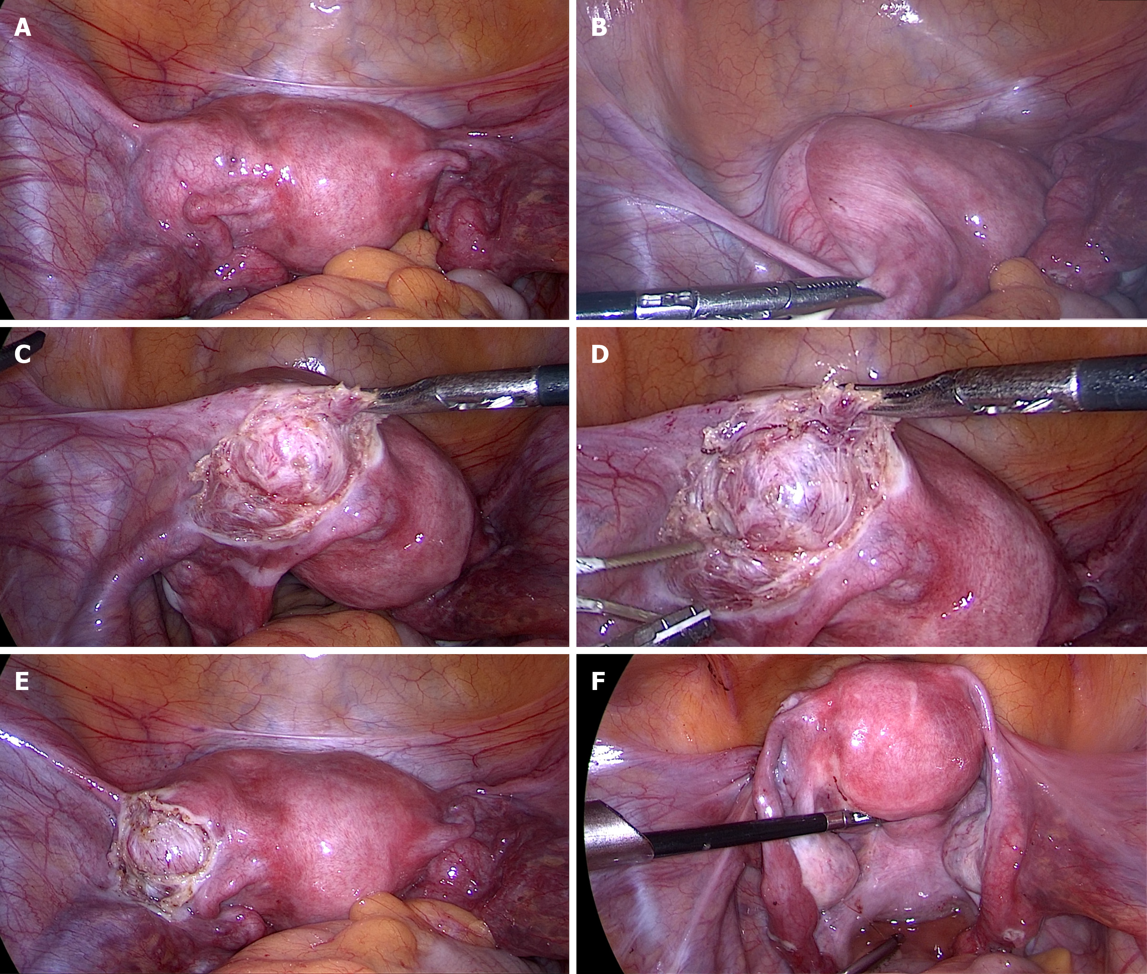
Figure 6 Internal genitalia of the patient showing a left accessory and cavitated uterine mass (A-F).
Uterus showing the accessory and cavitated uterine mass on the left anterior surface. Normal uterus and adnaexae were observed after the exeresis and peritonization.
- Citation: Hu YL, Wang A, Chen J. Diagnosis and laparoscopic excision of accessory cavitated uterine mass in a young woman: A case report. World J Clin Cases 2021; 9(30): 9122-9128
- URL: https://www.wjgnet.com/2307-8960/full/v9/i30/9122.htm
- DOI: https://dx.doi.org/10.12998/wjcc.v9.i30.9122


