
















©The Author(s) 2021.
World J Clin Cases. Oct 26, 2021; 9(30): 9114-9121
Published online Oct 26, 2021. doi: 10.12998/wjcc.v9.i30.9114
Published online Oct 26, 2021. doi: 10.12998/wjcc.v9.i30.9114
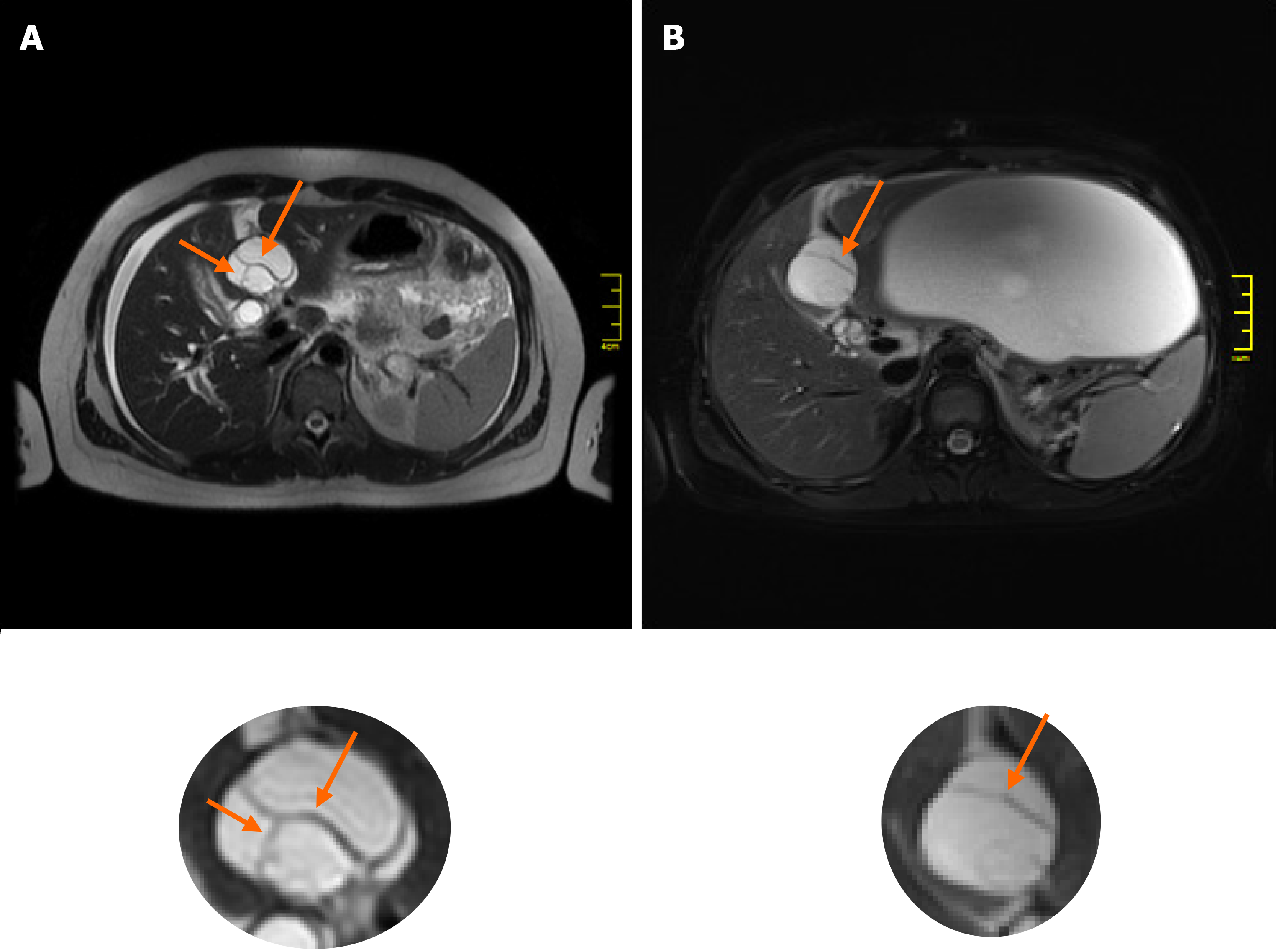
Figure 4 Changes in internal features of the cyst between magnetic resonance imaging studies.
Comparing the baseline magnetic resonance imaging (MRI) study (A) with the post-rupture MRI examination (B), the shape of the mucinous cystic neoplasm changed from oval to more circular. The internal architecture of its internal septations changed as demonstrated below (arrows).
- Citation: Kośnik A, Stadnik A, Szczepankiewicz B, Patkowski W, Wójcicki M. Spontaneous rupture of a mucinous cystic neoplasm of the liver resulting in a huge biloma in a pregnant woman: A case report. World J Clin Cases 2021; 9(30): 9114-9121
- URL: https://www.wjgnet.com/2307-8960/full/v9/i30/9114.htm
- DOI: https://dx.doi.org/10.12998/wjcc.v9.i30.9114