Copyright
©The Author(s) 2021.
World J Clin Cases. Oct 26, 2021; 9(30): 9114-9121
Published online Oct 26, 2021. doi: 10.12998/wjcc.v9.i30.9114
Published online Oct 26, 2021. doi: 10.12998/wjcc.v9.i30.9114
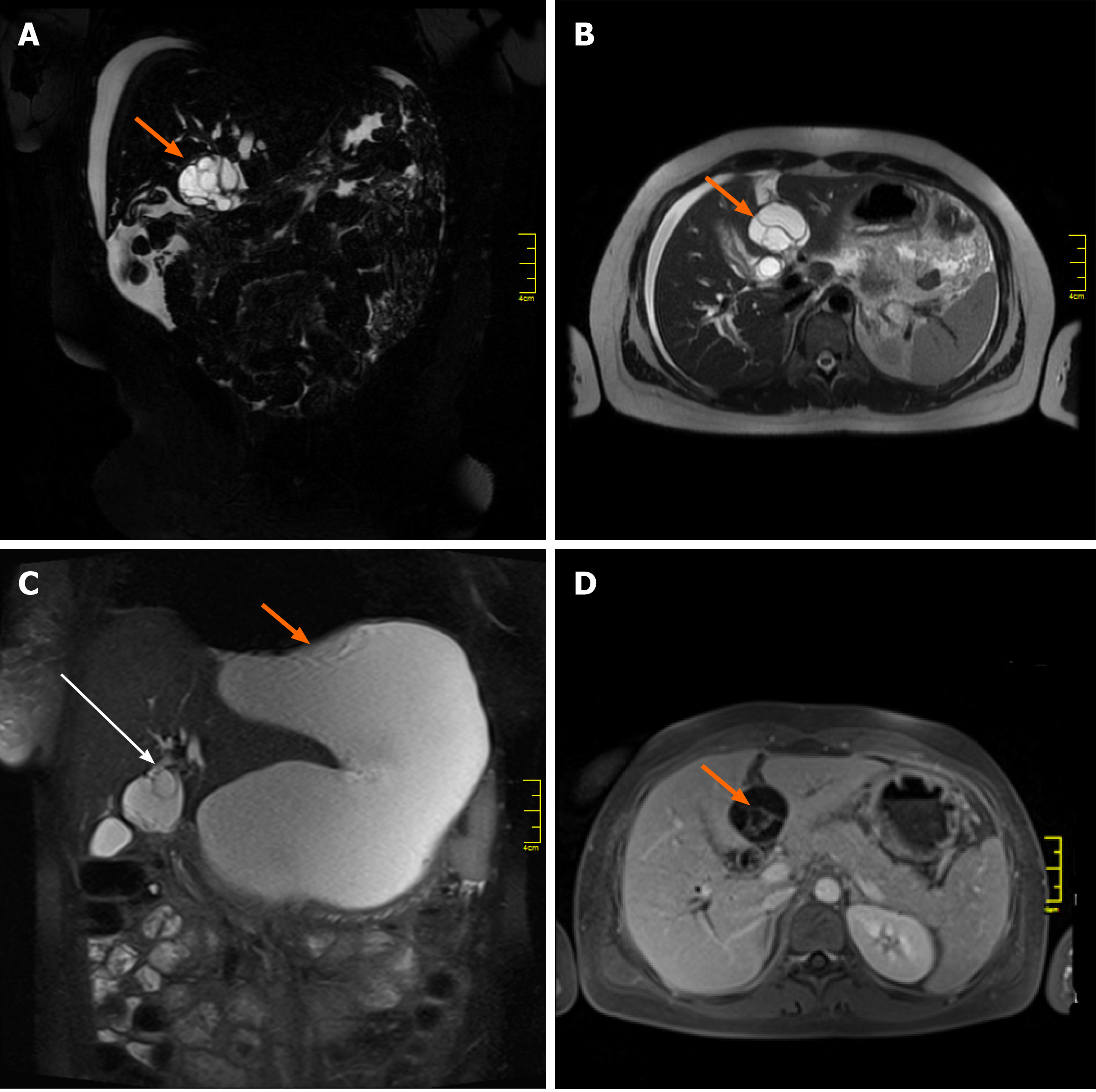
Figure 1 Magnetic resonance imaging studies of the cystic mass in the liver.
A, B: Initial magnetic resonance imaging (MRI) coronal and axial T2-weighted images showing a multilocular cystic mass (arrow), located between the left lateral segments of the liver (2/3) and segment 4; C: MRI T2-weighted coronal image demonstrating communication of the cystic mass with the left hepatic duct (long arrow) and a large fluid collection (short arrow) located anteriorly; D: MRI axial T1 image after intravenous gadolinium contrast administration, showing enhancement of internal septations (arrow) within the cystic liver mass.
- Citation: Kośnik A, Stadnik A, Szczepankiewicz B, Patkowski W, Wójcicki M. Spontaneous rupture of a mucinous cystic neoplasm of the liver resulting in a huge biloma in a pregnant woman: A case report. World J Clin Cases 2021; 9(30): 9114-9121
- URL: https://www.wjgnet.com/2307-8960/full/v9/i30/9114.htm
- DOI: https://dx.doi.org/10.12998/wjcc.v9.i30.9114


