Copyright
©The Author(s) 2021.
World J Clin Cases. Jan 26, 2021; 9(3): 758-763
Published online Jan 26, 2021. doi: 10.12998/wjcc.v9.i3.758
Published online Jan 26, 2021. doi: 10.12998/wjcc.v9.i3.758
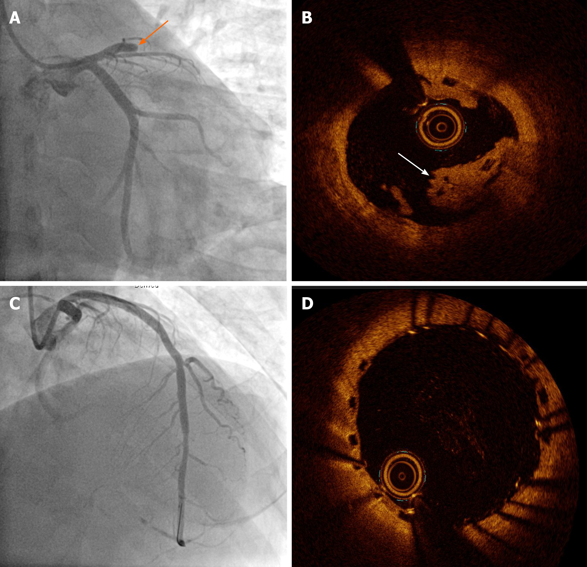
Figure 2 Very late scaffold thrombosis noted 20 mo ago.
A: Coronary angiography image showing total occlusion of the proximal left anterior descending artery; B: Optical coherence tomography image showing a red thrombus associated with disruption of the scaffold strut; C: Revascularization was successfully performed with the drug eluting stent (DES) covering the whole segment of the previous scaffold; D: Optical coherence tomography image showing optimal implantation of the DES on the previous bioresorbable vascular scaffold.
- Citation: Jang HG, Kim K, Park HW, Koh JS, Jeong YH, Park JR, Kang MG. Restenosis of a drug eluting stent on the previous bioresorbable vascular scaffold successfully treated with a drug-coated balloon: A case report. World J Clin Cases 2021; 9(3): 758-763
- URL: https://www.wjgnet.com/2307-8960/full/v9/i3/758.htm
- DOI: https://dx.doi.org/10.12998/wjcc.v9.i3.758


