Copyright
©The Author(s) 2021.
World J Clin Cases. Jan 26, 2021; 9(3): 659-665
Published online Jan 26, 2021. doi: 10.12998/wjcc.v9.i3.659
Published online Jan 26, 2021. doi: 10.12998/wjcc.v9.i3.659
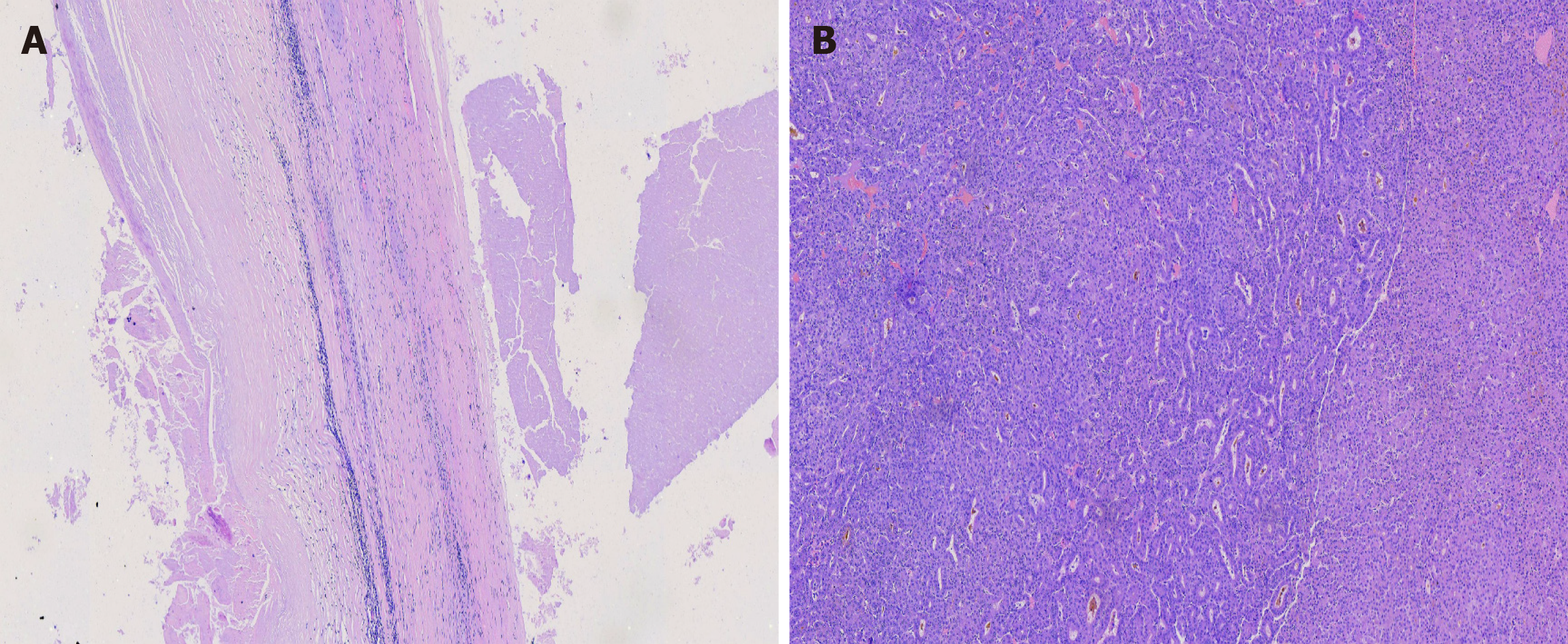
Figure 2 Microscopic histopathology images (× 100).
A: Section of specimens showed complete cyst wall structure, consistent with histopathological characteristics of cystic echinococcosis; B: Multiple highly differentiated cancer cells are found in the section specimens.
- Citation: Kalifu B, Meng Y, Maimaitinijiati Y, Ma ZG, Tian GL, Wang JG, Chen X. Radical resection of hepatic polycystic echinococcosis complicated with hepatocellular carcinoma: A case report. World J Clin Cases 2021; 9(3): 659-665
- URL: https://www.wjgnet.com/2307-8960/full/v9/i3/659.htm
- DOI: https://dx.doi.org/10.12998/wjcc.v9.i3.659


