Copyright
©The Author(s) 2021.
World J Clin Cases. Jan 26, 2021; 9(3): 573-580
Published online Jan 26, 2021. doi: 10.12998/wjcc.v9.i3.573
Published online Jan 26, 2021. doi: 10.12998/wjcc.v9.i3.573
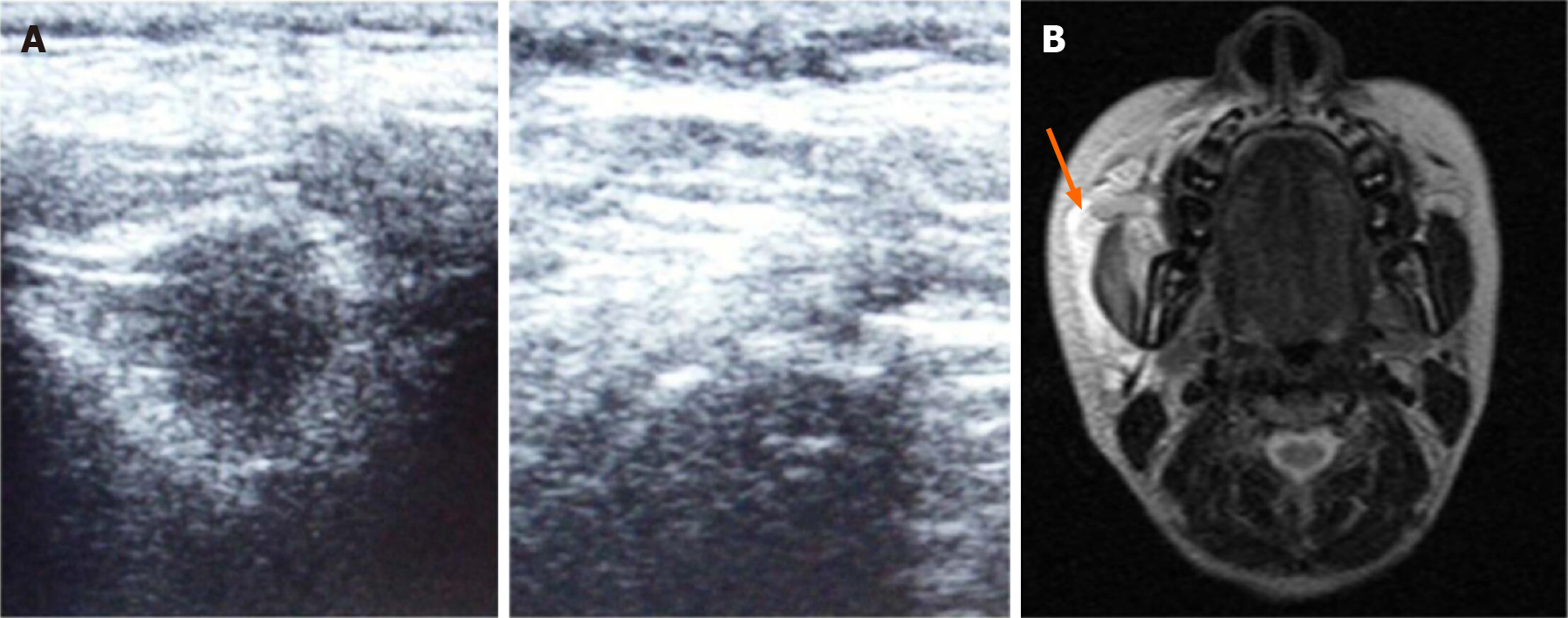
Figure 1 Comparison of ultrasonography and magnetic resonance images in case of the abscess.
A: The infected space showing the anechoic area with the surrounding wall. B: T2 axial-weighted magnetic resonance images with high signal intensity showing the involvement of buccal space.
- Citation: Ghali S, Katti G, Shahbaz S, Chitroda PK, V Anukriti, Divakar DD, Khan AA, Naik S, Al-Kheraif AA, Jhugroo C. Fascial space odontogenic infections: Ultrasonography as an alternative to magnetic resonance imaging. World J Clin Cases 2021; 9(3): 573-580
- URL: https://www.wjgnet.com/2307-8960/full/v9/i3/573.htm
- DOI: https://dx.doi.org/10.12998/wjcc.v9.i3.573


