Copyright
©The Author(s) 2021.
World J Clin Cases. Oct 16, 2021; 9(29): 8923-8931
Published online Oct 16, 2021. doi: 10.12998/wjcc.v9.i29.8923
Published online Oct 16, 2021. doi: 10.12998/wjcc.v9.i29.8923
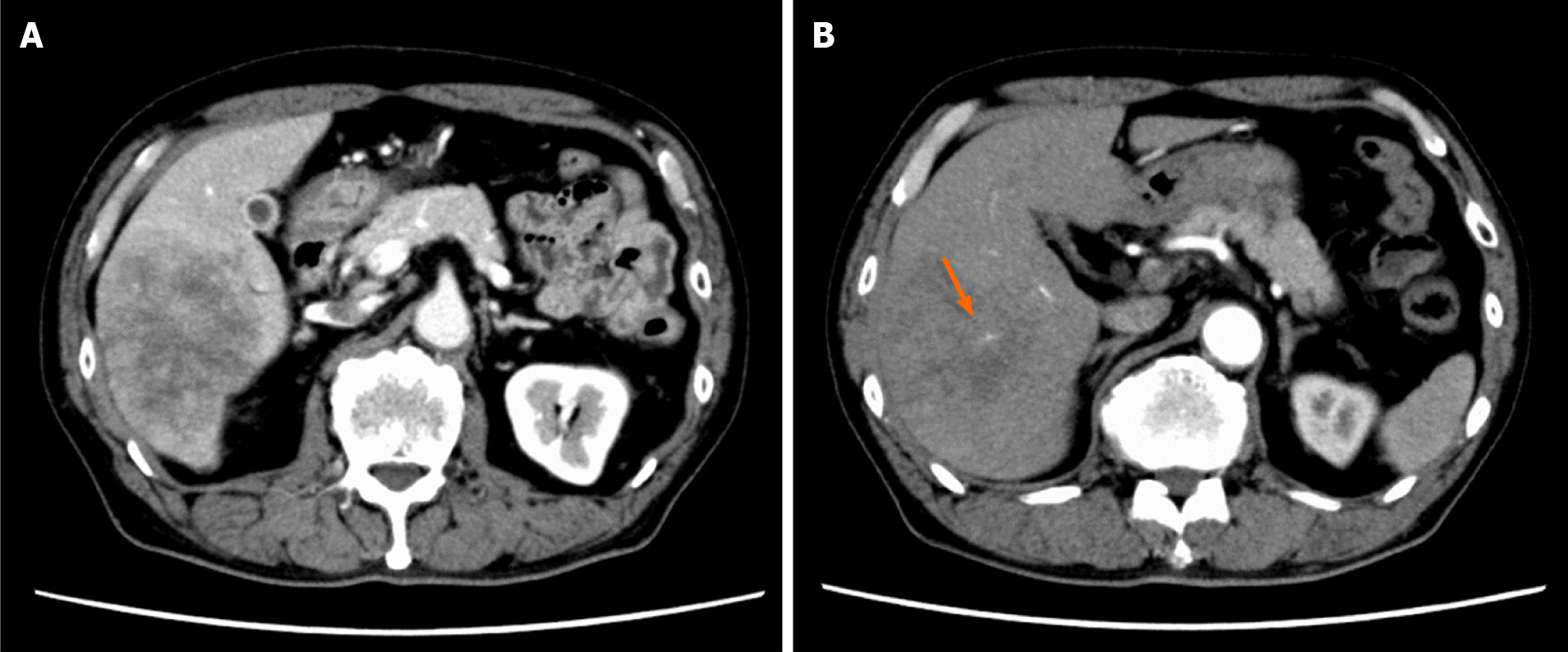
Figure 1 Image findings of contrast-enhanced dynamic-computed tomography.
A: Image showing a tumor with a diameter of approximately 8 cm in the posterior segment of the liver, which was weakly and gradually enhanced; B: Image showing an intratumoral artery in the arterial phase (arrow).
- Citation: Yonenaga Y, Yokoyama S. Isolated liver metastasis detected 11 years after the curative resection of rectal cancer: A case report. World J Clin Cases 2021; 9(29): 8923-8931
- URL: https://www.wjgnet.com/2307-8960/full/v9/i29/8923.htm
- DOI: https://dx.doi.org/10.12998/wjcc.v9.i29.8923


