Copyright
©The Author(s) 2021.
World J Clin Cases. Oct 16, 2021; 9(29): 8915-8922
Published online Oct 16, 2021. doi: 10.12998/wjcc.v9.i29.8915
Published online Oct 16, 2021. doi: 10.12998/wjcc.v9.i29.8915
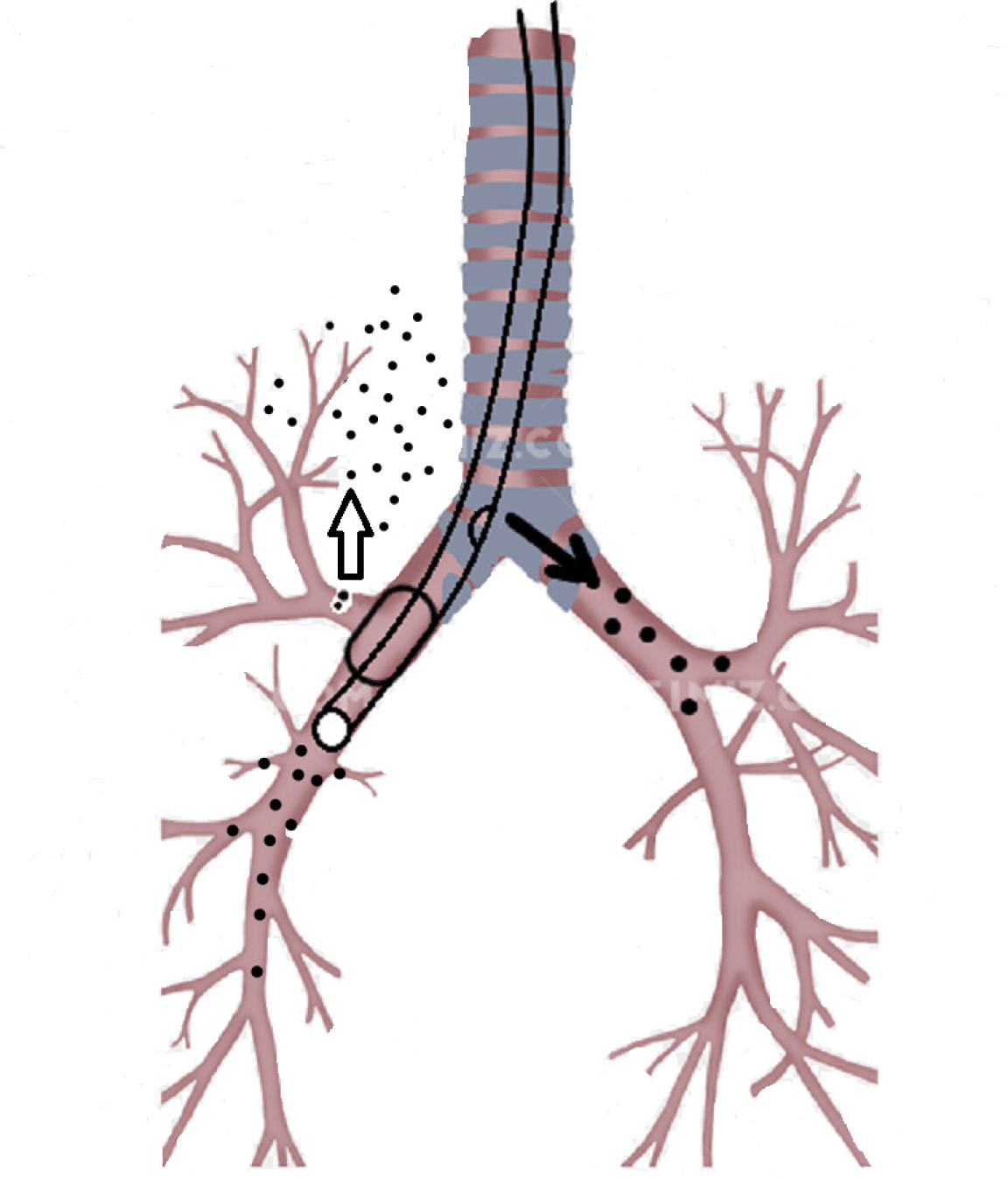
Figure 4 The modified tube was inserted under the guidance of a fiberoptic bronchoscope.
The incision in the tube was near the carina, which made it easy to ventilate the left lung through the incision. After the balloon was inflated, the balloon blocked the rupture in the bronchus. Using this method, the other right and left lung lobes were ventilated, thus avoiding atelectasis.
- Citation: Fan QM, Yang WG. Use of a modified tracheal tube in a child with traumatic bronchial rupture: A case report and review of literature. World J Clin Cases 2021; 9(29): 8915-8922
- URL: https://www.wjgnet.com/2307-8960/full/v9/i29/8915.htm
- DOI: https://dx.doi.org/10.12998/wjcc.v9.i29.8915


