Copyright
©The Author(s) 2021.
World J Clin Cases. Oct 16, 2021; 9(29): 8906-8914
Published online Oct 16, 2021. doi: 10.12998/wjcc.v9.i29.8906
Published online Oct 16, 2021. doi: 10.12998/wjcc.v9.i29.8906
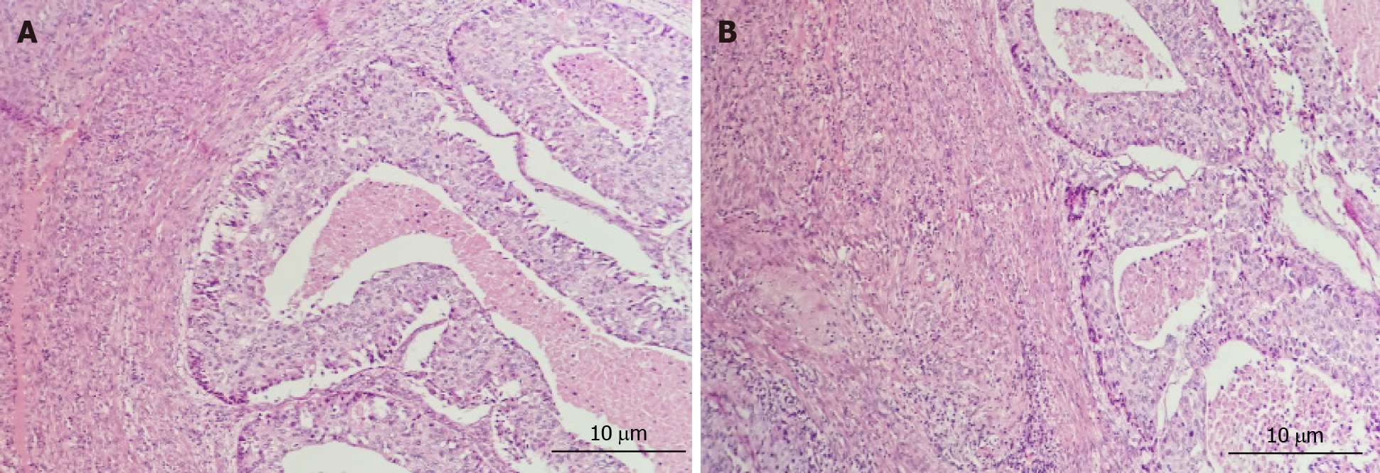
Figure 3 Pathological diagrams of endometrium.
Hematoxylin and eosin stain, × 200 magnification. A: Poorly differentiated endometrial carcinoma, increased lymphocytes infiltrate the cervical endometrium; B: Endometrioid carcinoma with squamous differentiation is not excepted, cytoplasm with eosinophilic keratinocytes, endometrial glandular differentiation.
- Citation: Yang L, Lin Y, Zhang XQ, Liu B, Wang JY. Acute pancreatitis with hypercalcemia caused by primary hyperparathyroidism associated with paraneoplastic syndrome: A case report and review of literature. World J Clin Cases 2021; 9(29): 8906-8914
- URL: https://www.wjgnet.com/2307-8960/full/v9/i29/8906.htm
- DOI: https://dx.doi.org/10.12998/wjcc.v9.i29.8906


