
















©The Author(s) 2021.
World J Clin Cases. Oct 16, 2021; 9(29): 8871-8878
Published online Oct 16, 2021. doi: 10.12998/wjcc.v9.i29.8871
Published online Oct 16, 2021. doi: 10.12998/wjcc.v9.i29.8871
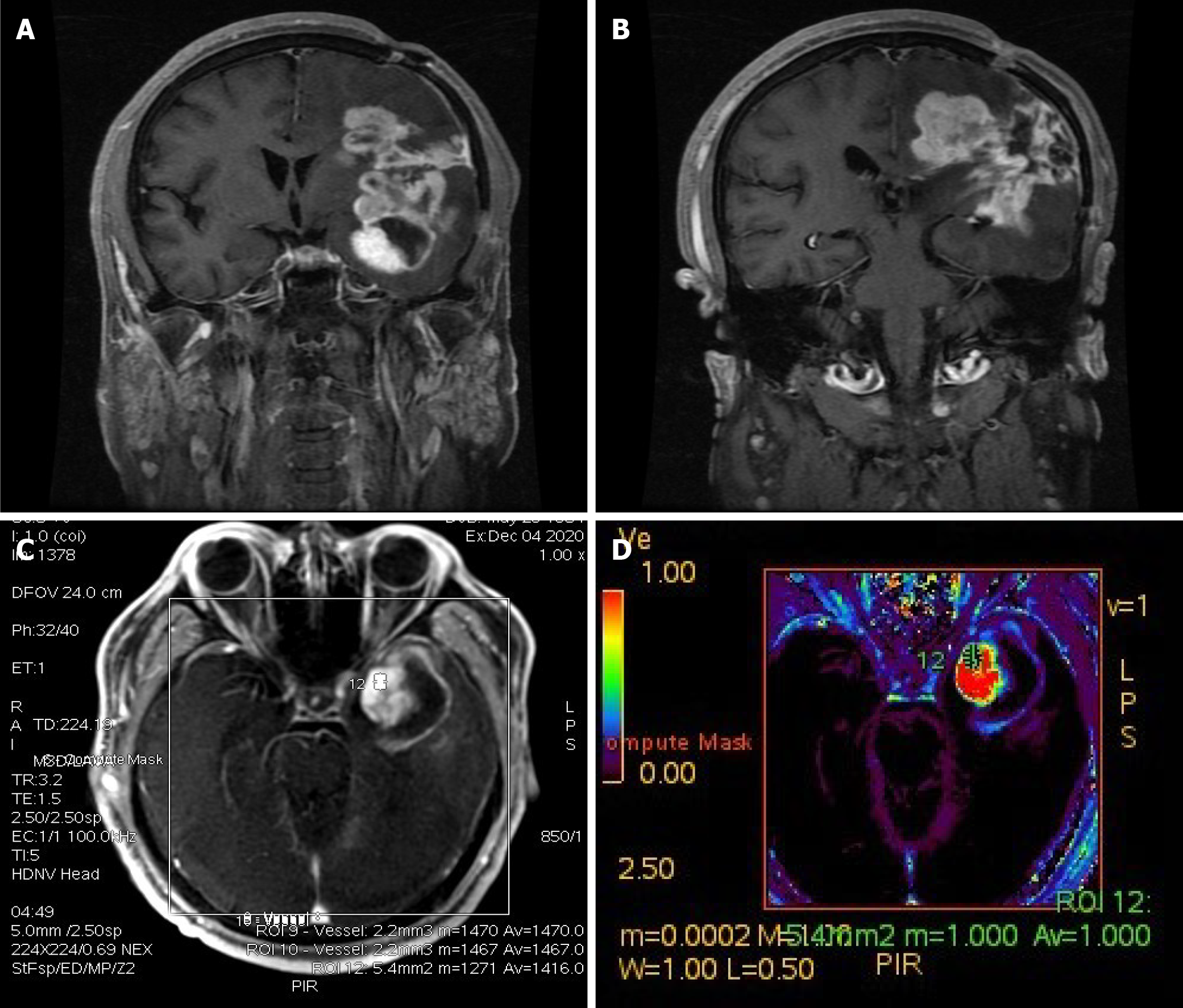
Figure 5 Imaging changes during patient follow-up.
A and B: Enhanced T1-weighted imaging 16 mo after craniotomy indicated enlargement of the lesion; C and D: Perfusion-weighted imaging of the enlarged part of the lesion (area marked by 12 in C) suggested that the enlarged lesion had high perfusion, and tumor recurrence was considered.
- Citation: Wang YY, Li ML, Zhang ZY, Ding JW, Xiao LF, Li WC, Wang L, Sun T. Primary intracranial synovial sarcoma with hemorrhage: A case report. World J Clin Cases 2021; 9(29): 8871-8878
- URL: https://www.wjgnet.com/2307-8960/full/v9/i29/8871.htm
- DOI: https://dx.doi.org/10.12998/wjcc.v9.i29.8871