Copyright
©The Author(s) 2021.
World J Clin Cases. Oct 16, 2021; 9(29): 8864-8870
Published online Oct 16, 2021. doi: 10.12998/wjcc.v9.i29.8864
Published online Oct 16, 2021. doi: 10.12998/wjcc.v9.i29.8864
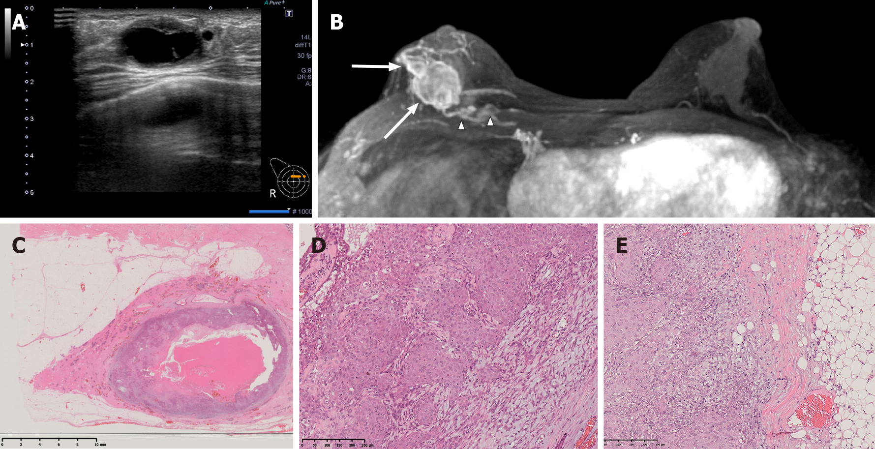
Figure 3 Imaging and pathology findings at the 2nd recurrence.
A: Ultrasonography shows a series of masses of up to 27 mm located on the right side of the previous surgical wound; B: Enhanced breast magnetic resonance imaging showed multiple masses up to 25 mm in the right inner-upper area (white arrow). The masses were cystic with thick walls, similar to the previous tumor, and were diagnosed as recurrent adenomyoepithelioma. Two 7-mm nodules were also found within the pectoralis major muscle (arrowhead); C: The tumor was a cystic lesion, and the cystic wall had nodules or irregular thickening. The cyst contained mucus (× 3 magnification); D: Biphasic proliferation of both inner epithelium with squamous metaplasia and outer spindle-shaped myoepithelium was seen (× 100 magnification). Both cell types showed prominent nuclear atypia and high mitotic counts that were especially prominent in the epithelial component. The histological findings were the same as those of the previous surgery; E: The tumor cells invaded the extramammary adipose tissue.
- Citation: Oda G, Nakagawa T, Mori M, Fujioka T, Onishi I. Adenomyoepithelioma of the breast with malignant transformation and repeated local recurrence: A case report. World J Clin Cases 2021; 9(29): 8864-8870
- URL: https://www.wjgnet.com/2307-8960/full/v9/i29/8864.htm
- DOI: https://dx.doi.org/10.12998/wjcc.v9.i29.8864


