Copyright
©The Author(s) 2021.
World J Clin Cases. Oct 16, 2021; 9(29): 8864-8870
Published online Oct 16, 2021. doi: 10.12998/wjcc.v9.i29.8864
Published online Oct 16, 2021. doi: 10.12998/wjcc.v9.i29.8864
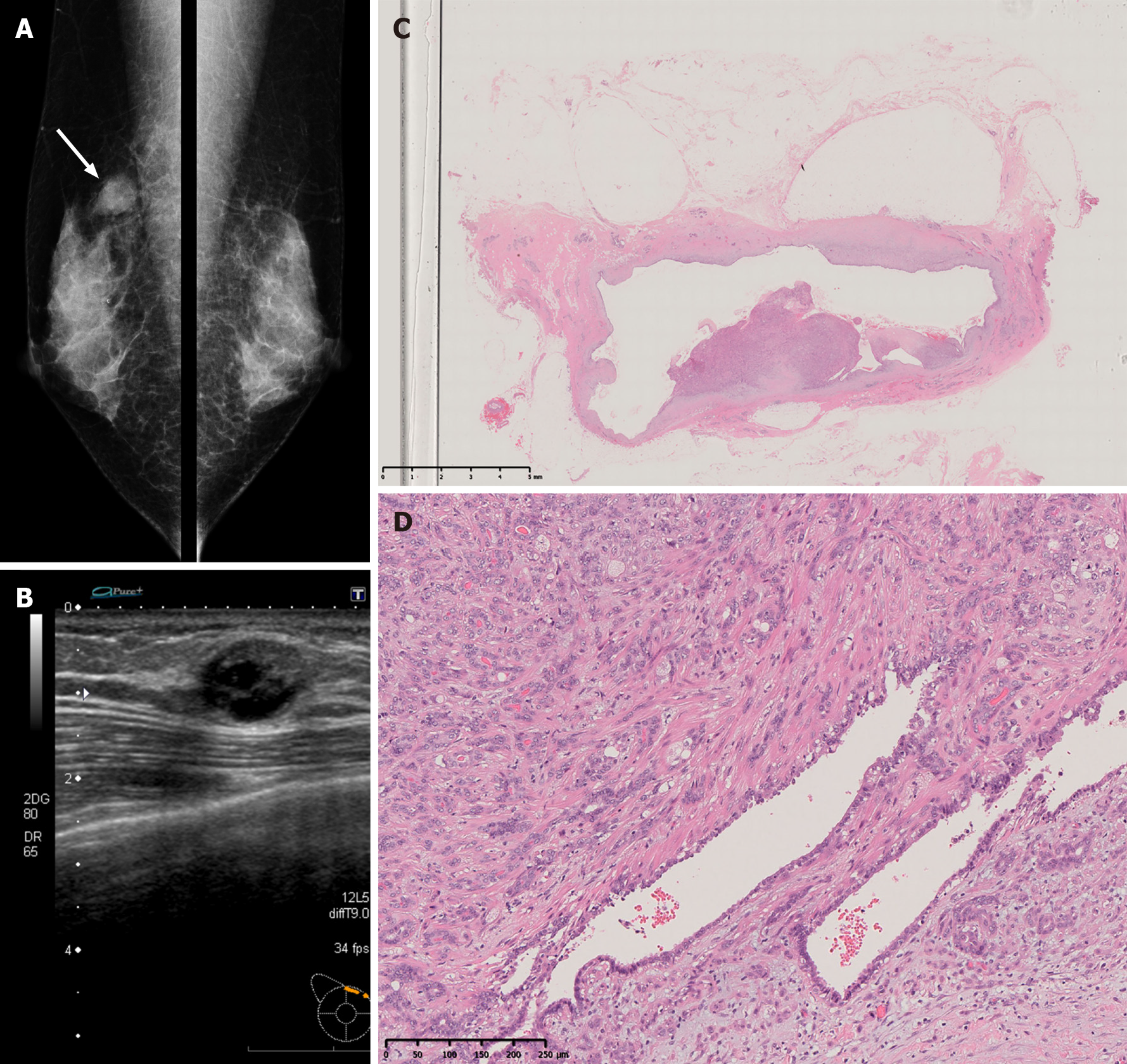
Figure 1 Imaging and pathology findings at the time of the initial surgery.
A: The mammogram showed an oval, smooth, well-defined, isodense mass in the upper right breast (white arrow); B: Ultrasonography showed a well-defined mass with cystic changes, measuring up to 16 mm, at the 2 o’clock position of the right breast; C: The tumor was a cystic lesion, and the cystic wall had nodules or irregular thickening (× 65 magnification); D: Round or spindle-shaped myoepithelium proliferating in and around the gland ducts was observed. High mitotic counts were prominent in the myoepithelial component (× 100 magnification).
- Citation: Oda G, Nakagawa T, Mori M, Fujioka T, Onishi I. Adenomyoepithelioma of the breast with malignant transformation and repeated local recurrence: A case report. World J Clin Cases 2021; 9(29): 8864-8870
- URL: https://www.wjgnet.com/2307-8960/full/v9/i29/8864.htm
- DOI: https://dx.doi.org/10.12998/wjcc.v9.i29.8864


