Copyright
©The Author(s) 2021.
World J Clin Cases. Oct 16, 2021; 9(29): 8782-8788
Published online Oct 16, 2021. doi: 10.12998/wjcc.v9.i29.8782
Published online Oct 16, 2021. doi: 10.12998/wjcc.v9.i29.8782
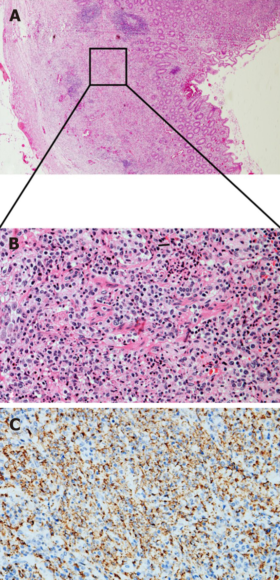
Figure 3 Pathological images of Samonellatyphi infection-related appendicitis.
A: Macrophage reactive hyperplasia in the submucosa is visible, and the normal lymphoid follicular structure disappears in the lamina propria (Hematoxylin-eosin staining, original magnification × 40); B: A magnification scope of Figure A, which shows massive macrophage reactive hyperplasia with a small amount of neutrophil and lymphocyte infiltration (Hematoxylin-eosin staining, original magnification × 400); C: Immunohistochemical staining for CD68 (Original magnification × 400).
- Citation: Zheng BH, Hao WM, Lin HC, Shang GG, Liu H, Ni XJ. Samonella typhi infection-related appendicitis: A case report. World J Clin Cases 2021; 9(29): 8782-8788
- URL: https://www.wjgnet.com/2307-8960/full/v9/i29/8782.htm
- DOI: https://dx.doi.org/10.12998/wjcc.v9.i29.8782


