Copyright
©The Author(s) 2021.
World J Clin Cases. Oct 6, 2021; 9(28): 8616-8626
Published online Oct 6, 2021. doi: 10.12998/wjcc.v9.i28.8616
Published online Oct 6, 2021. doi: 10.12998/wjcc.v9.i28.8616
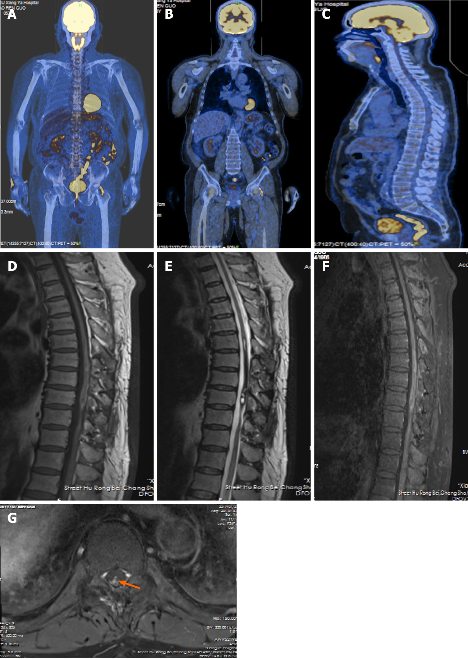
Figure 3 Postoperative positron emission tomography-computed tomography images and the latest review of magnetic resonance imaging.
A and B: Coronal positron emission tomography–computed tomography (PET-CT) images; C: Sagittal PET-CT image; D: Sagittal T1-weighted image (TIWI); E: Sagittal T2-weighted image; F: Sagittal TIWI with gadolinium enhancement; G: Axial TIWI with gadolinium enhancement at the T10 level. A-C: There were no abnormal regions with significant hypermetabolism in the whole body; D-G: The tiny residual (orange arrow) maintained stable. The heterogeneous enhancement of residual did not grow up, but the local edema of the spinal cord mitigated apparently.
- Citation: Liu ZQ, Liu C, Fu JX, He YQ, Wang Y, Huang TX. Primary intramedullary melanocytoma presenting with lower limbs, defecation, and erectile dysfunction: A case report and review of the literature. World J Clin Cases 2021; 9(28): 8616-8626
- URL: https://www.wjgnet.com/2307-8960/full/v9/i28/8616.htm
- DOI: https://dx.doi.org/10.12998/wjcc.v9.i28.8616


