Copyright
©The Author(s) 2021.
World J Clin Cases. Oct 6, 2021; 9(28): 8616-8626
Published online Oct 6, 2021. doi: 10.12998/wjcc.v9.i28.8616
Published online Oct 6, 2021. doi: 10.12998/wjcc.v9.i28.8616
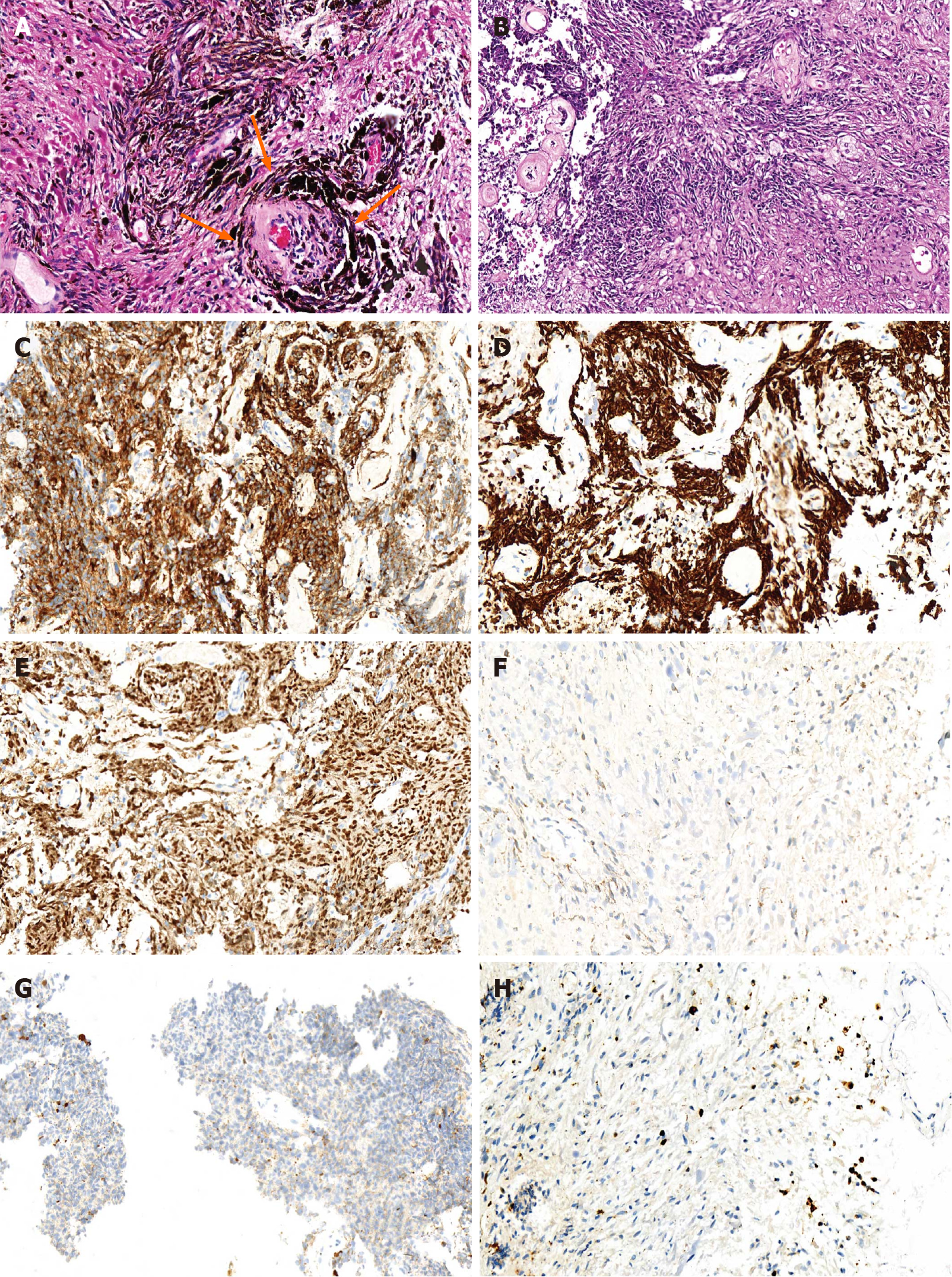
Figure 2 Postoperative pathology images showing the typical features of melanocytoma.
A: Hematoxylin and eosin (HE) staining (400 ×) revealed spindle and epithelioid cells containing melanin pigment in the cytoplasm assembled to form sheets, bundles, nests, or whorls surrounded by reticulin fibres (yellow arrow); B: HE staining (400 ×) after removing melanin pigment in the cytoplasm; C-H: Immunohistochemical staining (400 ×) demonstrated human melanoma black 45 (C), antimelanoma antibody (D), and sex determinant region Y box 10 protein (E) were positive. Besides, epithelial membrane antigen (F) and glial fibrillary acidic protein (G) were negative, and the proliferative index was only 3% (H).
- Citation: Liu ZQ, Liu C, Fu JX, He YQ, Wang Y, Huang TX. Primary intramedullary melanocytoma presenting with lower limbs, defecation, and erectile dysfunction: A case report and review of the literature. World J Clin Cases 2021; 9(28): 8616-8626
- URL: https://www.wjgnet.com/2307-8960/full/v9/i28/8616.htm
- DOI: https://dx.doi.org/10.12998/wjcc.v9.i28.8616


