Copyright
©The Author(s) 2021.
World J Clin Cases. Oct 6, 2021; 9(28): 8482-8491
Published online Oct 6, 2021. doi: 10.12998/wjcc.v9.i28.8482
Published online Oct 6, 2021. doi: 10.12998/wjcc.v9.i28.8482
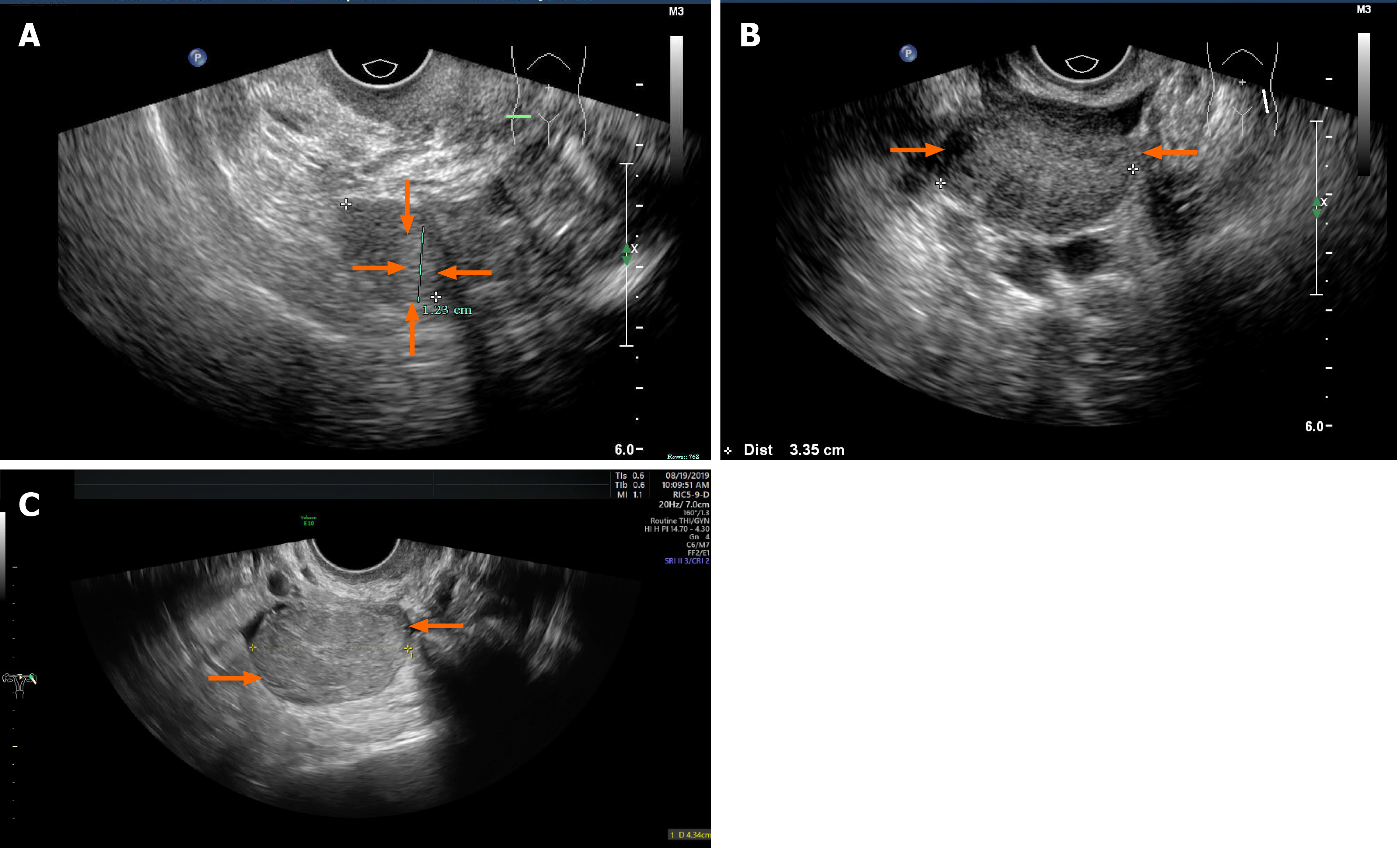
Figure 1 Transvaginal ultrasonography.
A: A slightly hyperechoic nodule with an unclear boundary in the right ovary (case 1); B: The left ovary showed a slightly hyperechoic mass with clear boundary (case 2); C: A solid, slightly hyperechoic mass with clear boundary in the left ovary (case 3).
- Citation: Zhu XD, Zhou LY, Jiang J, Jiang TA. Postmenopausal women with hyperandrogenemia: Three case reports. World J Clin Cases 2021; 9(28): 8482-8491
- URL: https://www.wjgnet.com/2307-8960/full/v9/i28/8482.htm
- DOI: https://dx.doi.org/10.12998/wjcc.v9.i28.8482


