Copyright
©The Author(s) 2021.
World J Clin Cases. Sep 26, 2021; 9(27): 8147-8156
Published online Sep 26, 2021. doi: 10.12998/wjcc.v9.i27.8147
Published online Sep 26, 2021. doi: 10.12998/wjcc.v9.i27.8147
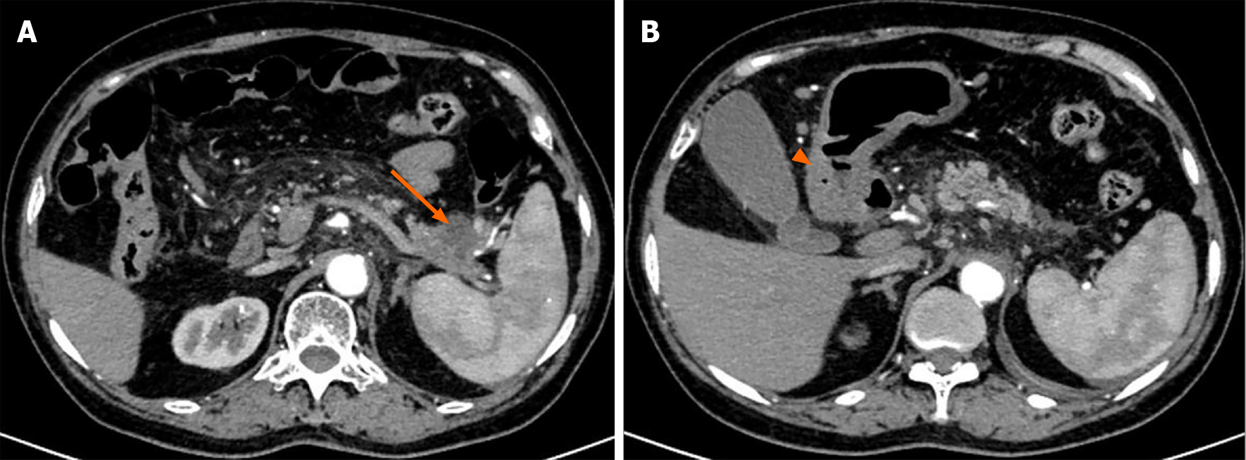
Figure 2 Enhanced upper abdominal computed tomography images.
A: A cystic solid mass measuring 3.5 cm × 3 cm × 2 cm in the cauda pancreas (orange arrow) surrounding the splenic vessels; B: Thickening of the antral wall and slight obstruction of the pylorus (orange arrowhead).
- Citation: Li K, Xu Y, Liu NB, Shi BM. Asymptomatic gastric adenomyoma and heterotopic pancreas in a patient with pancreatic cancer: A case report and review of the literature. World J Clin Cases 2021; 9(27): 8147-8156
- URL: https://www.wjgnet.com/2307-8960/full/v9/i27/8147.htm
- DOI: https://dx.doi.org/10.12998/wjcc.v9.i27.8147


