Copyright
©The Author(s) 2021.
World J Clin Cases. Sep 16, 2021; 9(26): 7954-7958
Published online Sep 16, 2021. doi: 10.12998/wjcc.v9.i26.7954
Published online Sep 16, 2021. doi: 10.12998/wjcc.v9.i26.7954
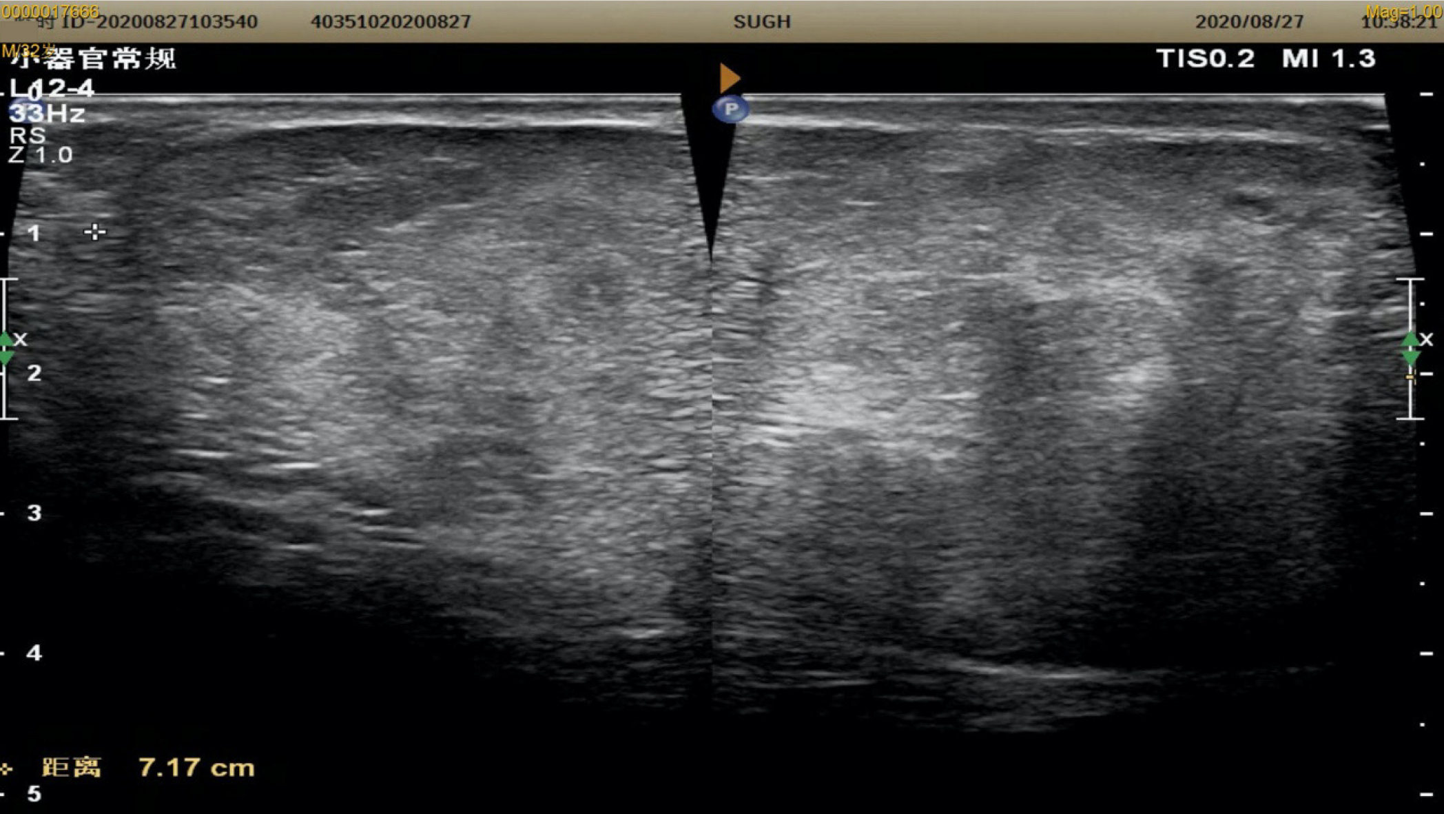
Figure 2 Colour Doppler ultrasound showing a hyperechoic mass under the skin of the left scrotum.
The mass measures about 72 mm × 64 mm × 41 mm, with clear boundaries, uneven internal echo, sinusoids, and strip-shaped blood flow signals; it is not connected to the abdominal cavity.
- Citation: Li SL, Zhang JW, Wu YQ, Lu KS, Zhu P, Wang XW. Subcutaneous angiolipoma in the scrotum: A case report. World J Clin Cases 2021; 9(26): 7954-7958
- URL: https://www.wjgnet.com/2307-8960/full/v9/i26/7954.htm
- DOI: https://dx.doi.org/10.12998/wjcc.v9.i26.7954


