Copyright
©The Author(s) 2021.
World J Clin Cases. Sep 16, 2021; 9(26): 7863-7869
Published online Sep 16, 2021. doi: 10.12998/wjcc.v9.i26.7863
Published online Sep 16, 2021. doi: 10.12998/wjcc.v9.i26.7863
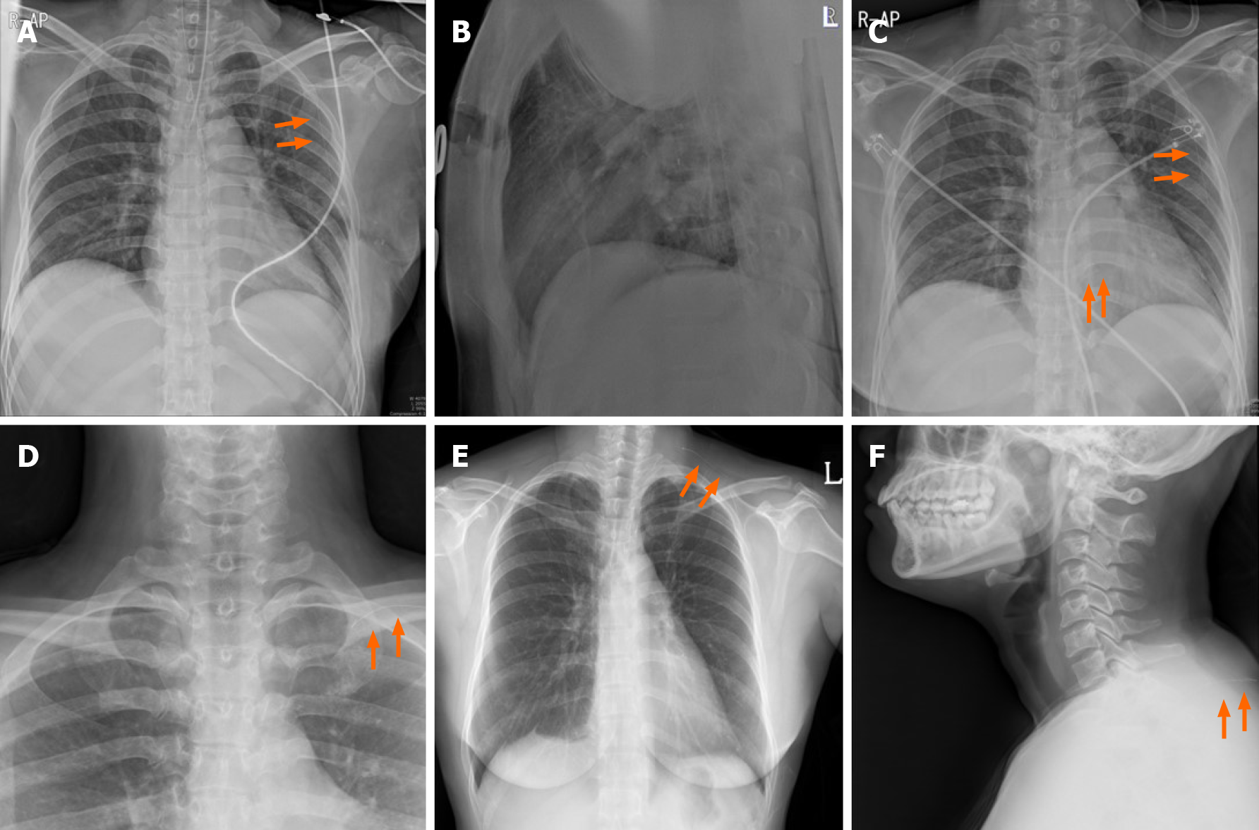
Figure 3 Serial simple X-ray image of the patient.
A and B: Intraoperative portable chest posteroanterior X-ray revealed the wire (orange arrows) to be located on midaxillary line-level longitudinally. But, it was not detected on lateral film; C: Recovery room portable chest posteroanterior X-ray revealed that the wire was located on midaxillary line-level longitudinally. This was the same finding in the intraoperative chest posteroanterior X-ray. No pneumothorax was seen; D: One day after the operation, a neck X-ray revealed the wire was located on the level of the clavicle; E and F: Two days after the operation, a serial simple X-ray revealed that the wire was located on a subcutaneous lesion of the back.
- Citation: Choi YJ. Migration of the localization wire to the back in patient with nonpalpable breast carcinoma: A case report. World J Clin Cases 2021; 9(26): 7863-7869
- URL: https://www.wjgnet.com/2307-8960/full/v9/i26/7863.htm
- DOI: https://dx.doi.org/10.12998/wjcc.v9.i26.7863


