Copyright
©The Author(s) 2021.
World J Clin Cases. Sep 16, 2021; 9(26): 7863-7869
Published online Sep 16, 2021. doi: 10.12998/wjcc.v9.i26.7863
Published online Sep 16, 2021. doi: 10.12998/wjcc.v9.i26.7863
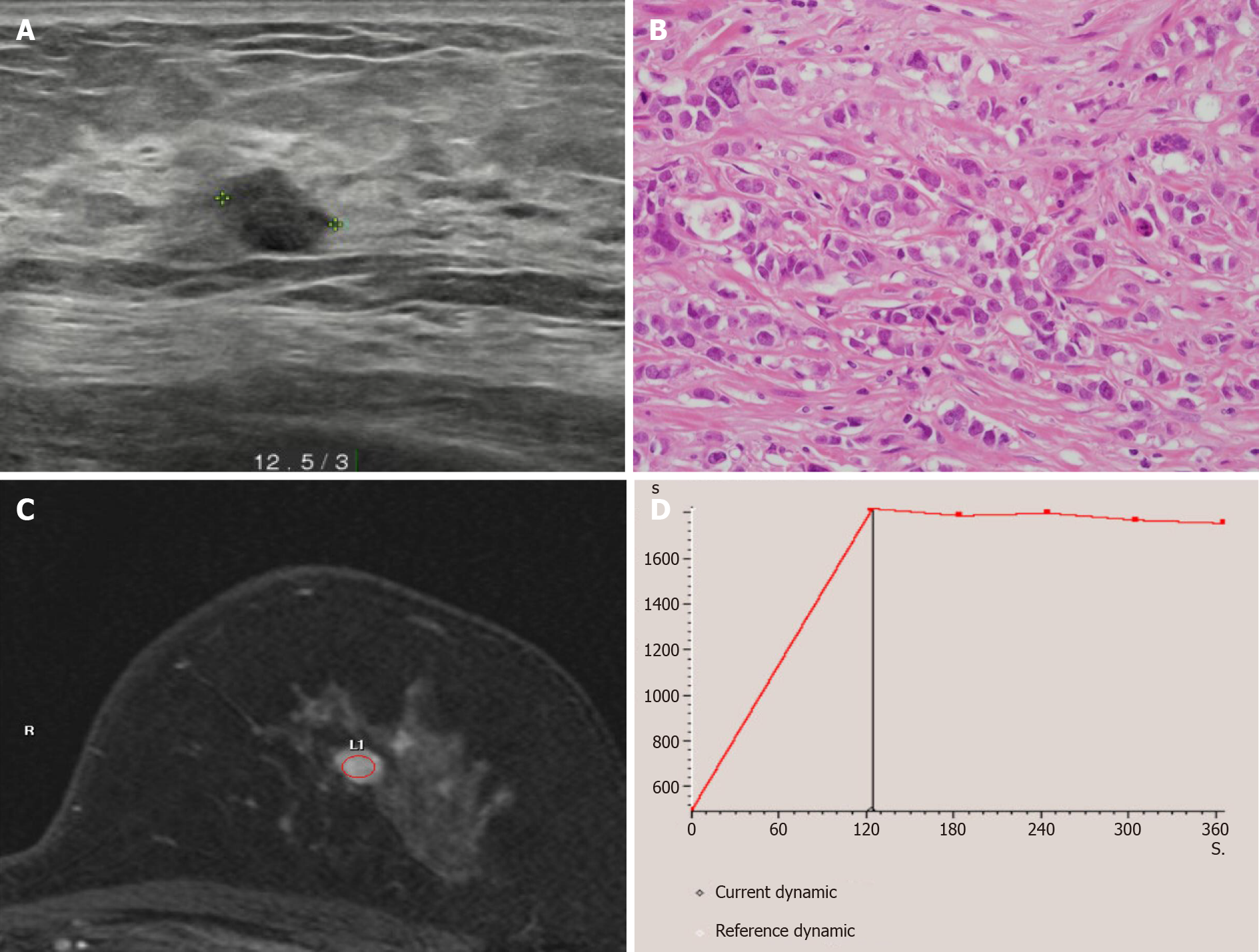
Figure 1 Radiologic findings of the left breast mass diagnosed as invasive ductal carcinoma.
A: Breast ultrasonography showed an 0.8 cm × 0.7 cm sized irregular hypoechoic mass located on the left 12:30 o’clock position at 3 cm distance from the left nipple; B: Breast core needle biopsy showed invasive ductal carcinoma with no special type (Haematoxylin and eosin staining × 400); C and D: Breast magnetic resonance imaging showed single enhancing mass on the left breast mass with a type II dynamic curve.
- Citation: Choi YJ. Migration of the localization wire to the back in patient with nonpalpable breast carcinoma: A case report. World J Clin Cases 2021; 9(26): 7863-7869
- URL: https://www.wjgnet.com/2307-8960/full/v9/i26/7863.htm
- DOI: https://dx.doi.org/10.12998/wjcc.v9.i26.7863


