Copyright
©The Author(s) 2021.
World J Clin Cases. Sep 16, 2021; 9(26): 7850-7856
Published online Sep 16, 2021. doi: 10.12998/wjcc.v9.i26.7850
Published online Sep 16, 2021. doi: 10.12998/wjcc.v9.i26.7850
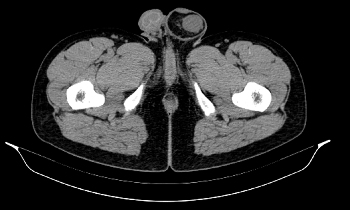
Figure 1 Pelvic computed tomography findings.
Pelvic computed tomography showed that intraperitoneal fat herniate in the left scrotum through widened left inguinal canal, and there is a mass appearing as soft tissue density in hernia contents with a size of approximately 2.5 cm × 1.3 cm × 3.0 cm.
- Citation: Liu JY, Li SQ, Yao SJ, Liu Q. Omental mass combined with indirect inguinal hernia leads to a scrotal mass: A case report. World J Clin Cases 2021; 9(26): 7850-7856
- URL: https://www.wjgnet.com/2307-8960/full/v9/i26/7850.htm
- DOI: https://dx.doi.org/10.12998/wjcc.v9.i26.7850


